Thank you for visiting nature.com. You are using a browser version with limited support for CSS. To obtain
the best experience, we recommend you use a more up to date browser (or turn off compatibility mode in
Internet Explorer). In the meantime, to ensure continued support, we are displaying the site without styles
and JavaScript.
Transparent tissues bring cells into focus for microscopy
Techniques that render tissues as clear as glass and swell them to several times their original size are giving unprecedented access to the inner workings of biological systems.
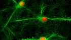
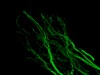
3D image of peripheral nerves (green) in a tissue-cleared 8-week-old human embryo.Credit: Alain Chédotal/Morgane Belle
In March, researchers in Japan mapped the cellular organization of the mouse brain in unprecedented detail.
Systems biologist Hiroki Ueda at the RIKEN Center for Biosystems Dynamics Research in Osaka, Japan, and his team created an atlas of the mouse brain using a technique called CUBIC-X, in which they chemically labelled every cell in the brain, then rendered the organ crystal-clear while also expanding its size tenfold1. From there, they used sophisticated imaging techniques to compile a comprehensive 3D neuronal survey — of some 72 million cells in all, Ueda says. The resulting atlas reduces the brain to a compact database of cellular addresses, which the team used to explore changes in various brain regions during development. Moving forward, the atlas could drive deeper explorations of brain structures that control behaviours such as the sleep–wake cycle.
CUBIC-X is just one component in a growing toolbox of such methods, which exploit readily available chemicals to provide researchers with a window not just into the brain, but into virtually every organ in the body. Some are tissue-clearing methods that make opaque tissues transparent, whereas others complement tissue clearing with a proportional size increase that exposes molecular details to conventional microscopy. The choice comes down to the scientific question. There are many ways to achieve similar ends, and users should investigate the strengths and limitations of different methods before deciding which to use.
Mind readers
The hunger for tissue-clearing techniques originated with neuroscientists, who were frustrated by their limited ability to trace the snaking routes of axons and dendrites in the brain.
Such studies conventionally involve serial imaging of thin sections of labelled brain tissue, followed by computational reconstruction into 3D. But the process is slow: imaging circuits in a single mouse brain can entail weeks in front of a microscope. And the resulting maps are only as good as the input data. “Most times, you are only sampling a handful of slices, and this is not very efficient for reconstruction,” says Viviana Gradinaru, a neuroscientist at the California Institute of Technology in Pasadena. “And cutting can damage the tissue surface and edges in a way that prevents you from realigning them.”
A better approach would be to make the tissue transparent and then image it intact. But only in the past few decades have molecular reagents, genetic strategies and imaging techniques advanced far enough to make that possible.
When it comes to illuminating the brain’s interior, lipids are public enemy number one. As light passing through an aqueous solution encounters a lipid surface, the change in refractive index causes it to bend and scatter. “Think about Jell-O [a jelly]: it’s made mainly of proteins and it’s translucent,” says Gradinaru. “But if you add cream to the Jell-O, it becomes opaque. That cream is made of lipids.” Cell and organelle membranes are made mainly of lipids, as are the myelin sheaths enveloping axons. Clearing the brain entails eliminating these molecules, while physically stabilizing the molecules that remain behind.
German anatomist Werner Spalteholz first demonstrated a strategy for clearing opaque tissues in 1911, using chemical solvent treatments that eliminated light-scattering biomolecules. But that method was incompatible with today’s fluorescent reagents, and damaging to tissue structures.
In 2011, Hans-Ulrich Dodt, a brain-imaging specialist at the Vienna University of Technology; Ali Ertürk, now at the University of Munich, Germany; and Frank Bradke at the German Center for Neurodegenerative Diseases in Bonn described one of the first modern clearing techniques. Called 3DISCO, the method is a spiritual descendant of Spalteholz’s protocol, using a gentler cocktail of chemical solvents that dissolve lipids while preserving cellular structures and dehydrating and hardening the specimen into a clear framework that retains the tissue’s original structure2. “We think that the solvent-based methods are the most reliable in terms of reproducibility and cost — the first time you do it, it works,” says Alain Chédotal, a developmental neuroscientist at the INSERM Vision Institute in Paris. Solvent-based methods are generally best suited for use with fluorescently tagged antibodies as the reporter molecules, because genetically expressed fluorescent proteins tend to yield a weakened signal or be denatured by such treatments. But a new variant of 3DISCO from Ertürk’s team overcomes that problem. vDISCO uses dye-labelled ‘nanobodies’ to boost the signal from fluorescent proteins in solvent-cleared tissues — an approach the team used to clear and image intact mice (see go.nature.com/2tk6hr3).
Another widely used tissue-clearing option is CLARITY, which Gradinaru helped to develop as a graduate student in neuroscientist Karl Deisseroth’s lab at Stanford University in California in 20133. The Deisseroth lab makes extensive use of fluorescent proteins in its neuroscience research, and sought a more ‘naturalistic’ clearing approach that minimized damage to biomolecules of interest. CLARITY uses detergent to eliminate lipids, while reinforcing the tissue infrastructure with polymers that form a water-based hydrogel. “All of those monomers hold hands with each other and they lock in the proteins as well,” explains Gradinaru, “and then you can come in with that gentle detergent to take the lipids out.” Early versions of CLARITY were technically challenging, requiring an electrical field to actively drive out detergent-encapsulated lipids. But Gradinaru subsequently devised a simpler alternative that perfuses the animal’s vasculature with clearing solution to achieve the same effect.
For the CUBIC family of techniques, Ueda developed yet another approach4. CUBIC exploits ‘hydrophilic’ chemicals that draw water into the fixed specimen while pushing dissolved lipids out. As with CLARITY, the method preserves the structure and function of fluorescent proteins while clarifying the sample.
Body of evidence
Clearing methods allow researchers to tackle neuroscience questions that were previously out of reach. Chédotal, for instance, is using 3DISCO to explore the intact wiring of the visual system. “From the eye, you have the output of ganglion cells that project to maybe 30 different parts of the brain,” he says. “How these axons find these different targets is completely unknown.”
But the methods aren’t limited to the brain. “Much to our surprise, most of the rodent organs turned transparent within a few days,” says Gradinaru of her work in developing perfusion-based CLARITY. “It resulted in the whole body being cleared except the skin and bones,” she says. When it comes to tissue clearing, bone poses a particular challenge, Gradinaru says, because calcium continues to reflect light even after conventional clearing. But additional treatments can eliminate this problem. Ueda, for instance, has shown that EDTA, a commonly used laboratory chemical, efficiently removes calcium from bone. Gradinaru’s team has also developed a bone-clearing CLARITY variant.
Such methods make it possible to perform whole-body imaging of intact specimens, and researchers have applied them to track rare populations of stem cells or tumour metastases, and even to trace the developing vasculature and peripheral nervous system in first-trimester human embryos. As samples get bigger, however, microscopy can become a bottleneck, and researchers must balance research goals with practicality. Light-sheet microscopy is one popular solution. “You’re scanning a plane of light through the tissue rather than a point, and that greatly accelerates the imaging,” says Deisseroth. So, too, is the physical shrinkage that accompanies certain clearing methods. In 3DISCO, dehydration can reduce sample size by up to 50%. “That means we can image a whole human embryo in 3D in one shot,” says Chédotal.
Still, the envelope can be pushed only so far. Chédotal, who has cleared human brains, has had to content himself with imaging regions measuring a few cubic centimetres, roughly 1% of the organ. “I could make a whole cow transparent,” he says. “But I wouldn’t be able to image it, so what’s the point?”
The big picture
Clearing is a valuable starting point for tissue imaging, but to make out fine molecular details, researchers also needs the means to ‘zoom in’.
Conventional light microscopes are unable to distinguish molecules separated by less than the ‘diffraction limit’ of light, around 200 nanometres — a problem that led to the development of technically sophisticated super-resolution microscopy approaches. Ed Boyden, a neurobiologist at the Massachusetts Institute of Technology (MIT) in Cambridge, was joking when he initially proposed an alternative approach — ‘blowing up’ the brain. But soon he saw real potential in the idea. “All the proteins in the cell are jam-packed together, and if we expand them apart from each other, maybe we could see them better,” he says.
Enjoying our latest content?
Log in or create an account to continue
Access the most recent journalism from Nature's award-winning team
Explore the latest features & opinion covering groundbreaking research


 ‘Invisible’ mice reveal anatomical secrets
 See-through brains clarify connections
 Blown-up brains reveal nanoscale details