Thank you for visiting nature.com. You are using a browser version with limited support for CSS. To obtain
the best experience, we recommend you use a more up to date browser (or turn off compatibility mode in
Internet Explorer). In the meantime, to ensure continued support, we are displaying the site without styles
and JavaScript.
In the age of immunotherapy, cancer biologists are relying on a new generation of tools to learn how the interplay between tumours and immune cells shapes the course of disease.
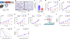
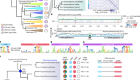
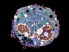
The microenvironment surrounding a breast-cancer tumour (blue) contains different types of immune cells, such as T cells and B cells. Credit: Akoya Biosystems
Although he is trained as a pathologist, Sean Bendall has become something of a map-maker of late, using cutting-edge protein-mapping technology to chart the treacherous landscape of tumour tissues.
Created with Michael Angelo, Bendall’s colleague at Stanford University in California, this technology is providing rich molecular profiles of both cancer cells and their immediate neighbours — most notably, the immune cells in and around the tumour. Bendall is already seeing evidence that these maps could help clinicians to decide on the appropriate treatment for some people. In a 2018 study1, Bendall and Angelo applied their technique, called multiplexed ion beam imaging (MIBI), to tumour specimens from people undergoing standard chemotherapy for triple-negative breast cancer, which is a particularly aggressive type of tumour. “We identified an immune type that was highly predictive of long-term disease-free survival,” Bendall says.
It’s easy to think of tumours in stark, binary terms, composed of cells that are either abnormal or healthy. But the reality is messier: cancer cells interact extensively with immune cells, blood vessels and supporting connective tissues. This tumour ‘microenvironment’ can profoundly affect both the characteristics of the disease and a person’s response to treatment, particularly immunotherapy — a type of treatment that helps the immune system to fight cancer. The microenvironment, for example, can determine whether nearby immune cells are switched ‘on’ or ‘off’, or even whether they can access the tumour at all. “The tumour is not just a homogenous bag of cells,” says Dana Pe’er, a computational biologist at the Memorial Sloan-Kettering Cancer Center in New York. “It’s actually an organ. It’s just a badly malformed organ.”
Armed with tumour-mapping technologies, such as Bendall’s, as well as other approaches that can generate detailed censuses of vast numbers of individual cells based on gene expression or protein content, researchers are now dissecting the structure and function of the tumour microenvironment. These tools are so new that many researchers are still getting the hang of how best to use them. But the resulting insights could help to usher in a new era of tumour profiling — one that takes a ‘big picture’ view of cellular ecosystems, such as the number and type of immune cells present, rather than focusing exclusively on individual genetic variants.
“These are features that are really going to impact treatment,” says Miriam Merad, a medical oncologist at the Icahn School of Medicine at Mount Sinai in New York City.
A cellular census
The idea that interplay between the tumour and the immune system might shape a person’s disease is not new — immunologist Wolf Fridman has been studying such interactions for 50 years, based on early observations of the response of immune cells called T cells in leukaemia. “I rapidly got the conviction that the location and organization of the immune reaction is very important,” says Fridman, who is now an emeritus professor at the Medical School Paris Descartes. However, the broader impact of these interactions didn’t become a major focus for oncologists until about ten years ago, with the emergence of powerful immunotherapy strategies.
Pathologists can get some indication of the immune-cell composition of a tumour with conventional techniques such as haematoxylin and eosin (H&E) staining and immunohistochemistry (IHC). IHC, which uses enzyme-tagged antibodies that recognize specific molecular features in a tissue specimen, has proved particularly informative in the context of the various drugs known as checkpoint inhibitors. This class of immunotherapy agent blocks specific signalling proteins that can otherwise prevent local immune cells from mounting an effective attack on the tumour, and IHC can reveal the presence of these proteins. “If you don’t have a certain level of checkpoint-protein expression in your lung tumour, you basically don’t get a response,” says Bendall. However, many people that do express these proteins still fail to respond to checkpoint therapy, and researchers are hunting for other immune features that might more clearly predict whether the treatment will be effective. But IHC is not an optimal strategy for finding these, because it can only profile a handful of molecular markers at a time.
MIBI-generated molecular profiles of tumour and immune cells. Cells are coloured to show either the distance from the tumour border (left) or the expression of genetic markers (right).Credit: Ref. 1
Mass cytometry by time-of-flight, or CyTOF, can profile tumour cells on a much larger scale. Instead of coupling antibodies to dyes or fluorescent labels, as in conventional flow cytometry, CyTOF uses antibodies linked to metal isotopes to label large numbers of dissociated cells from a tumour specimen. These isotope tags are then rapidly profiled with a mass spectrometer, which can detect and quantify dozens of different markers in parallel for each cell.
In 2017, Merad and her colleagues applied CyTOF to lung adenocarcinoma — the most common form of lung cancer — using as many as 40 different tagged antibodies2. Their data revealed how newly emerging tumours move quickly to quell a person’s immunity by recruiting immune-suppressing regulatory T cells and impeding the migration of ‘effector’ cells such as tumour-slaying natural killer cells. “Even at this early stage, when they start to become malignant, they have to start getting rid of these effector cells,” says Merad. “These results suggest that reversing immunosuppression very early in tumour development could halt progression and, hopefully, metastasis.”
Tallying transcripts
CyTOF requires prior knowledge of cell-type-specific markers so that researchers can select the appropriate antibodies, limiting its utility in discovery-based research. And Pe’er notes that CyTOF offers little direct information on biological function, indicating only the presence or absence of cellular markers.
Transcriptome analysis techniques such as RNA sequencing (RNA-seq) can overcome some of these limitations. RNA-seq employs high-throughput sequencing platforms to characterize and quantify vast numbers of protein-coding messenger RNA transcripts. This offers a direct window onto which genes are turned off and on in a given cell, revealing the biological activities that take place in a tumour. Researchers have been applying such techniques for more than a decade, but much of this has been ‘bulk’ analysis — profiling every cell in the tumour at once. That approach can offer useful insights, but it also glosses over any variation between cells, notes Aviv Regev, a computational biologist at the Broad Institute in Cambridge, Massachusetts.
Regev’s lab helped to develop a method called Drop-seq, in which individual cells are encapsulated and prepared for sequencing in individual lipid droplets. During this process, each cell’s RNA is assigned a distinct genetic barcode, making it straightforward to determine which transcripts originate from the same cell. “We get very comprehensive profiles — both the variety of cells, and the variety of molecules inside the cells,” says Regev. Crucially, Drop-seq and other single-cell RNA-seq methods require no prior knowledge of the genes of interest, and the gene-expression profiles can even be used to reconstruct interactions between cells, as Regev demonstrated last year for melanoma tumours3. “We found that malignant cells actually can assume a specific state where they form what are called ‘cold niches’, where you don’t have T cells,” Regev says. Such tumour regions could prove more resistant to immunotherapy.
Single-cell RNA-seq is relatively straightforward to implement, with several commercial instruments available. But the technique still poses some important challenges. Perhaps the most significant is that tumour specimens must be shuttled directly from the operating theatre to the laboratory, with only a brief layover in the pathologist’s lab. “Single-cell approaches are only really informative on fresh cells, because the RNA is very quickly degraded when we freeze and thaw cells,” says Merad.
However, the technology can uncover cell types and cell states that researchers might otherwise not have known to look for, and reveal nuances in immune-cell function that transcend the simplistic categories often used to sort cells. “There’s much more variation and many more subsets than we imagined,” says Pe’er. “We used to like to rank cells as ‘good’ and ‘bad’, in terms of pro- or antitumour, but the system is much more complex, with most cells co-expressing both good and bad programs.”
Lay of the land
For all their power, CyTOF and RNA-seq come with a serious trade-off: destruction of the tumour to access individual cells. This erases the spatial details that can be invaluable for understanding interactions between malignant cells and their microenvironment. Methods such as IHC and H&E staining capture such details, but can reveal only limited information in a single experiment, confounding efforts to assemble a comprehensive portrait of the microenvironment. “I’ve literally watched pathologists in our department putting slides on top of one another to look at multiple features at the same time,” says Bendall. A pair of imaging techniques developed in 2014 can deliver this kind of rich spatial data without the need for stacking and squinting.
The first, imaging mass cytometry, was developed by Bernd Bodenmiller of the University of Zurich, Switzerland, and his colleagues4. As with conventional CyTOF, samples are labelled with metal isotope-tagged antibodies, but in Bodenmiller’s technique, the labelling is performed on intact tissue rather than on dissociated cells. The specimens are then scanned with a laser that disrupts the tissue and releases the isotope labels, which are analysed with CyTOF. This enables simultaneous measurement of dozens of markers at subcellular resolution across an intact sample with a level of detail that greatly exceeds IHC. “Imaging mass cytometry has an orders-of-magnitude higher dynamic range, and that really brings a much more quantitative aspect to the analysis of immune markers,” says Bodenmiller.
The second technique is MIBI, developed by Angelo and Bendall while the two were working in immunologist Garry Nolan’s lab at Stanford5. MIBI uses isotopically labelled antibodies and a scanning ion beam that subsequently liberates those labels from the specimen, but it uses a different kind of mass spectrometer for isotope analysis. Angelo and Bendall have continued to refine this platform to improve its speed and ease of use, as highlighted in their 2018 work with triple-negative breast cancer1. “Instead of taking almost a day to acquire one tiny picture, we ended up acquiring millimetre-square images from an entire cohort of about 40 patients over the course of about a week and a half,” says Bendall.
Although they have been used only in a handful of published studies, MIBI and imaging mass cytometry are already highlighting the value of knowing where cells dwell in the tumour microenvironment. “Everyone says that tumours are highly heterogeneous and random systems, but we’ve found a lot of structure and cell–cell interactions that are not random,” says Bodenmiller.
Enjoying our latest content?
Log in or create an account to continue
Access the most recent journalism from Nature's award-winning team
Explore the latest features & opinion covering groundbreaking research