Figure 1

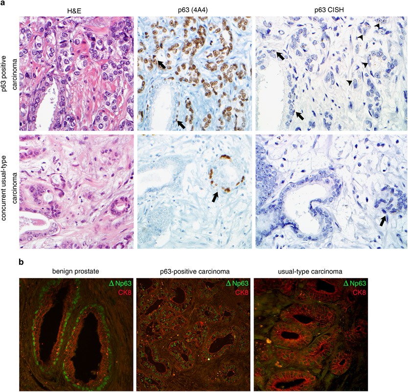
p63-expressing prostate cancers are positive for p63 mRNA by chromogenic in situ hybridization (CISH) and the ΔNp63 isoform by immunohistochemistry. (a) p63-expressing prostate carcinoma (top row) expresses p63 protein in a non-basal cell distribution (using the 4A4 antibody which detects both the ΔNp63 and TAp63 isoforms, × 400 magnification) as well as p63 mRNA by CISH (arrowheads), at levels similar to or higher than surrounding benign basal cells (arrow, × 630 magnification). The concurrent usual-type adenocarcinoma in this case (bottom row) does not express p63 protein or mRNA; however, nearby benign basal cells are positive for both (arrows). (b) Dual ΔNp63 and CK8 staining in prostatic tissues. These markers are expressed in separate compartments in benign prostatic tissue, with basal cells expressing ΔNp63 and luminal cells expressing CK8 (left panel). In p63-expressing tumors, these two markers are expressed in the same cells (middle panel). In contrast, usual-type acinar carcinomas express CK8 and are negative for ΔNp63. All images at × 400 magnification.
