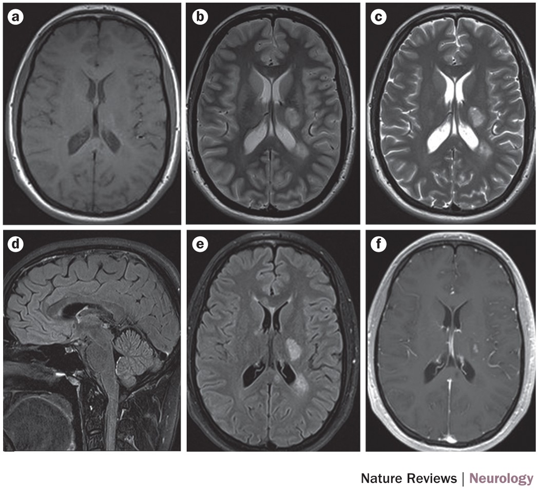
Figure 1: Standardized brain MRI protocol to evaluate patients in whom multiple sclerosis is clinically suspected.

a | Pre-contrast, b | axial T1-weighted and c | dual-echo T2-weighted sequences, followed by d | contrast-enhanced sagittal, e | axial 2D T2-weighted fluid-attenuated inversion recovery (FLAIR) and f | axial T1-weighted sequences. With this strategy, there is no penalty in terms of total acquisition time, and it ensures a minimum delay of 5 min between gadolinium injection and acquisition of the T1-weighted sequence.
