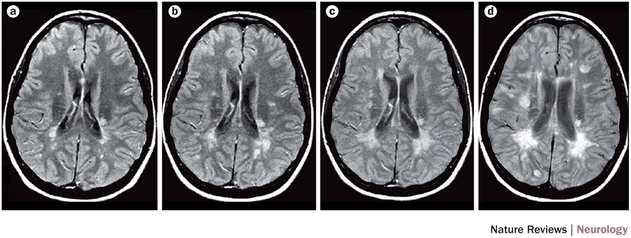
Figure 1: Serial MRI in a patient with relapsing–remitting multiple sclerosis.

Proton-density weighted MRI scans obtained at a | baseline, and b | 1 year, c | 2 years and d | 3 years later. Disease progression can clearly be seen in the form of new and enlarging focal lesions over time, shown here as hyperintensities (white spots).
