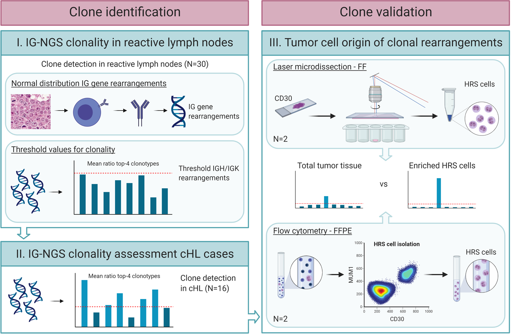
Fig. 1: Graphical summary of the workflow for clone identification and validation in classic Hodgkin lymphoma.

For clone identification in classic Hodgkin lymphoma (cHL), first the relative frequencies of the most-abundant clonotypes in non-malignant reactive lymph nodes (N = 30) were determined by next-generation sequencing of immunoglobulin gene rearrangements (IG-NGS) (Supplementary Fig. S1). Here, clonality analysis was performed in duplicate, followed by calculation of the mean ratio of the top-4 overlapping clonotypes identified in duplicate in each sample, for each of the immunoglobulin heavy chain (IGH) and immunoglobulin kappa light chain (IGK) targets, including both complete and incomplete IG gene rearrangements. Based on the maximum ratio of the overlapping clonotypes in the reactive lymph nodes, a threshold value for clonality was defined for each of the targets. The established threshold values were used to define clonality in the 16 cHL cases. For clone validation, laser microdissection and flow cytometry were used to enrich the Hodgkin Reed-Sternberg (HRS) cells from the tumor-tissue specimen of two cHL cases, and these HRS cell fractions were subjected to IG-NGS clonality analysis to validate these as neoplastic clones.
