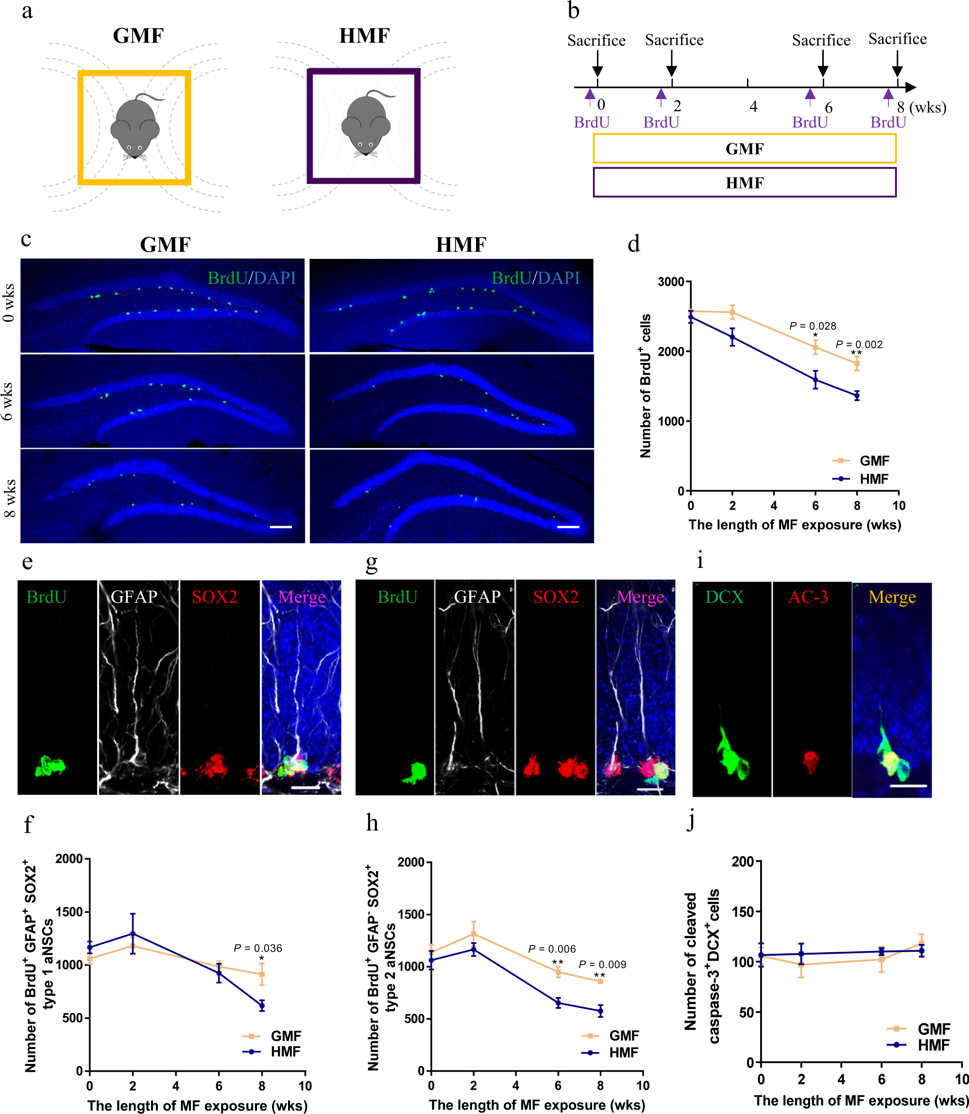
Fig. 1: Long-term HMF exposure decreased the proliferation of aNSCs in the DG of mice.
From: Long-term exposure to a hypomagnetic field attenuates adult hippocampal neurogenesis and cognition

a Schematic diagram of magnetic field exposure to adult mice. b Experiments timeline for cell proliferation analysis during HMF or GMF exposure. c Representative images of BrdU+ cells in the DG after 2-h BrdU pulse labeling at 0-, 6-, 8-weeks of GMF- or HMF-exposure. Scale bar = 200 μm. d Quantification of numbers of BrdU+ cells in GMF- and HMF-exposed mice (GMF versus HMF, two-way ANOVA, F(1, 23) = 24.96, P < 0.0001). e Representative images of BrdU+GFAP+Sox2+ type 1 aNSCs in the adult DG. Scale bar = 20 μm. f Quantification of numbers of BrdU+GFAP+Sox2+ type 1 aNSCs in GMF- and HMF-exposed mice (GMF versus HMF, two-way ANOVA, F(1, 20) = 0.2978, P = 0.5913). g Representative images of BrdU+GFAP-Sox2+ type 2 aNSCs in the adult DG. Scale bar = 20 μm. h Quantification of numbers of BrdU+GFAP−Sox2+ type 2 aNSCs in GMF- and HMF-exposed mice (GMF versus HMF, two-way ANOVA, F(1, 21) = 14.81, P = 0.0009). i Representative images of brain section stained with DCX and activated caspase-3 (AC-3) in the adult DG. Scale bar = 20 μm. j Quantification of numbers of AC-3+DCX+ cells in GMF- and HMF-exposed mice (GMF versus HMF, two-way ANOVA, F(1, 24) = 0.186, P = 0.67). GMF, n = 4 mice, HMF, n = 4 mice. Data are presented as mean ± SEM. All data were analyzed by two-way ANOVA, and the two-tailed unpaired t test for two-group comparisons at each time point.
