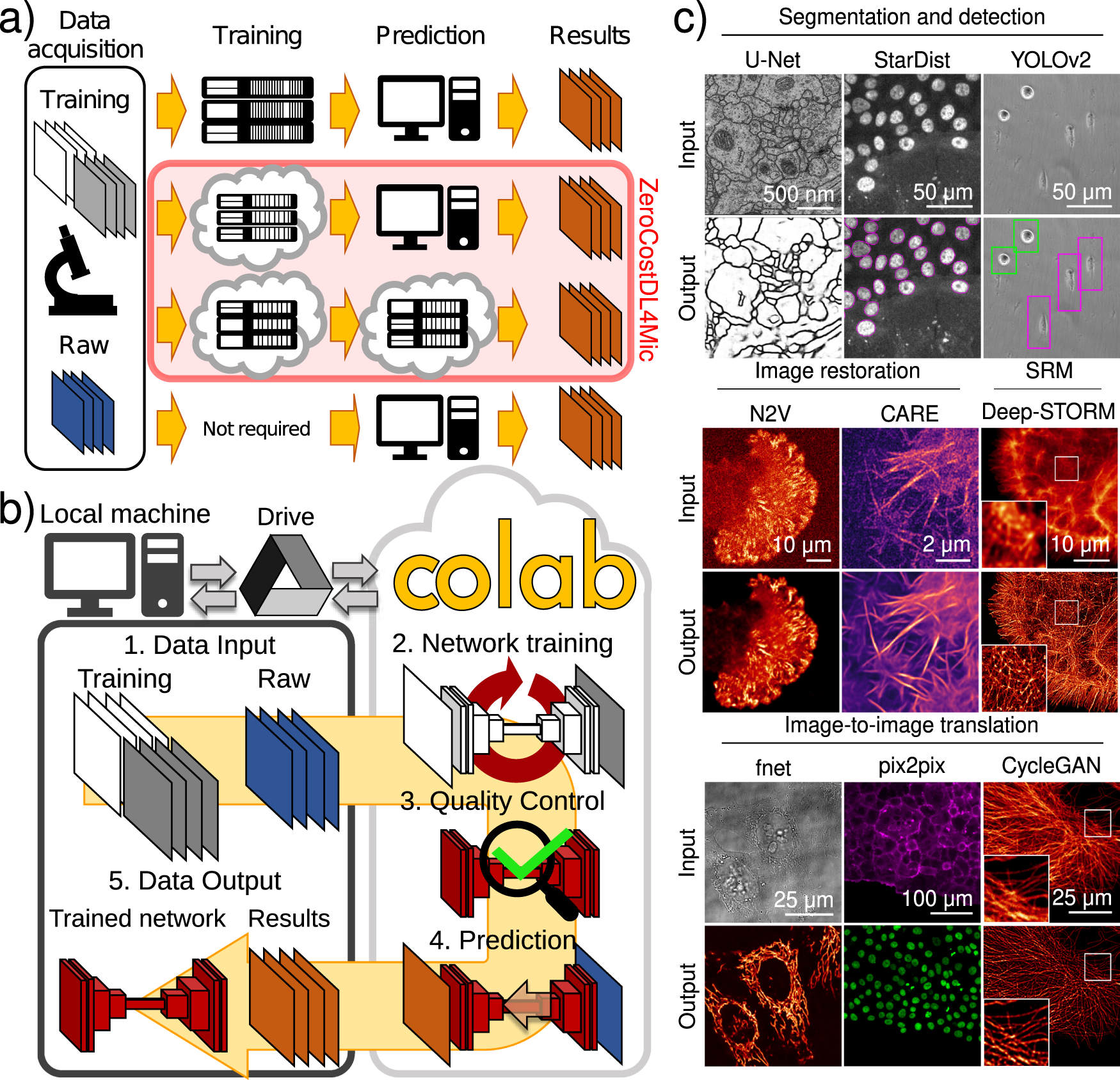
Fig. 1: Using DL for microscopy.
From: Democratising deep learning for microscopy with ZeroCostDL4Mic

a Paths to exploiting DL. Training on local servers and inference on local machines (or servers) (first row), cloud-based training and local inference (second row), cloud-based training and inference (third row) and pretrained networks on standard machines (fourth row). b Overview of ZeroCostDL4Mic. The workflow of ZeroCostDL4Mic, featuring data transfer through Google Drive, training, quality control and prediction via Google Colab. After running a network, trained models, quality control and prediction results can then be downloaded to the user’s machine. c Overview of the bioimage analysis tasks currently implemented within the ZeroCostDL4Mic platform. Datasets from top left to bottom right: U-Net—ISBI 2012 Neuronal Segmentation Dataset78,79, StarDist—nuclear marker (SiR-DNA) in DCIS.COM cells, YOLOv2—bright field in MDA-MB-231 cells, N2V—actin label (paxillin-GFP) in U-251-glioma cells, CARE—actin label Lifeact-RFP in DCIS.COM cells, Deep-STORM—actin-labelled glial cell, fnet—bright-field and mitochondrial label TOM20-Alexa Fluor 594 in HeLa cells, pix2pix—actin label Lifeact-RFP and nuclear labels in DCIS.COM cells, CycleGAN—tubulin label in U2OS cells. All datasets are available through Zenodo (see “Data availability”) or as indicated in the GitHub repository.
