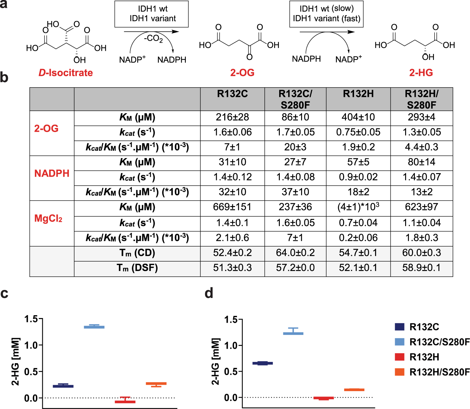
Fig. 1: The IDH1 S280F substitution increases the efficiency of active site variants.

a IDH1 variant catalysed oxidation of isocitrate and reduction of 2-OG to 2-HG. b Kinetic parameters for IDH1 variants (400 nM) from non-linear regression curve fits (standard error of the mean, n = 3). Conditions: 100 mM Tris, 10 mM MgCl2, 0.2 mM DTT, 0.005%v/v Tween 20, and 0.1 mg/mL BSA (pH 8.0). Shaded: Melting temperatures (Tms) of IDH1 variants measured by differential scanning fluorimetry (DSF, 3 µM enzyme) or circular dichroism (CD, 0.2 mg/mL enzyme); λ: 215 nm. Conditions: DSF (20 mM Tris, 100 mM NaCl, pH 7.4); CD (10 mM sodium phosphate, pH 8.0). See Supplementary Information for details. c 2-HG formation from 2-OG catalysed by IDH1 variants as measured by 1H NMR (700 MHz; error bars: standard errors of the mean, n = 3 independent replicates of 1H time course experiments). Conditions: 500 nM enzyme, 10 mM MgCl2, 1.5 mM 2-OG, 1.5 mM NADPH; incubation time: 12 min. Source data are provided as a Source Data file. d 2-HG formation from isocitrate catalysed by IDH1 variants as measured by NMR (700 MHz; standard error of the mean, n = 3 independent replicates of 1H time course experiments). Conditions: 750 nM enzyme, 10 mM MgCl2, 3 mM DL-isocitrate, 1.5 mM NADP+; incubation time: 9 min. Buffer: 50 mM Tris-d11, 100 mM NaCl, 10 mM MgCl2, and 10% D2O, pH 7.5. Source data are provided as a Source Data file.
