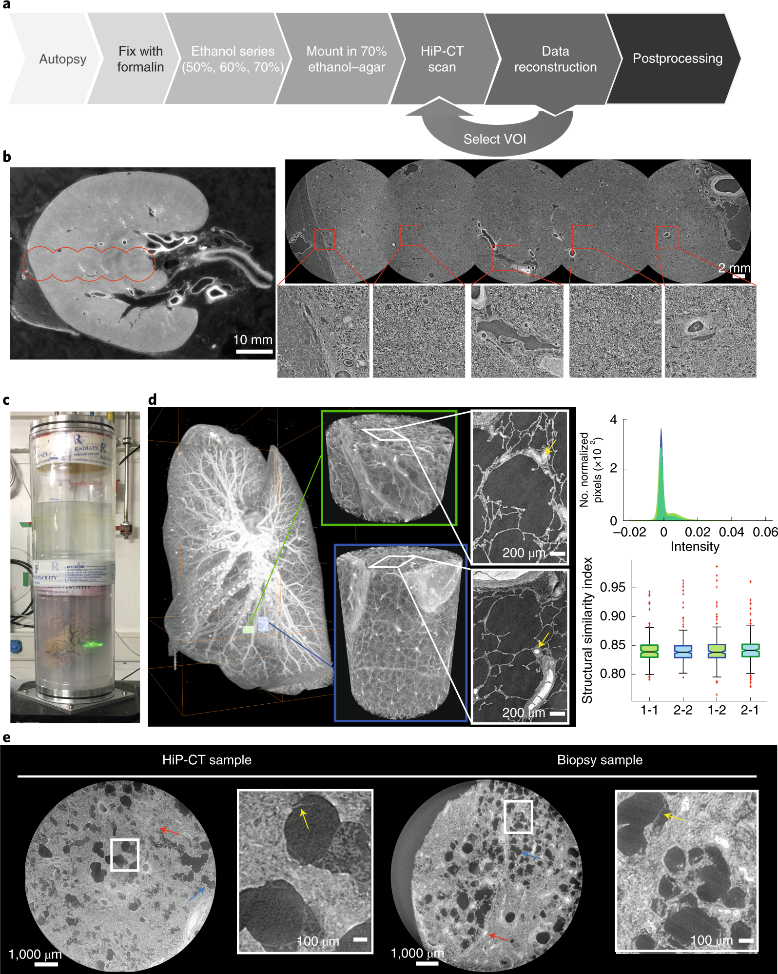
Fig. 1: A HiP-CT pipeline for multiscale 3D imaging from whole-organ to cellular resolution within large intact soft tissue samples.

a, Flow chart of HiP-CT sample preparation and imaging procedure; the ability to select specific higher-resolution scan regions based on lower-resolution scans provides hierarchical tissue structure images in a data-efficient manner. b, Left, 2D image slice (25 µm per voxel) showing the location of a series of regions of 2.5 µm per voxel that transect the organ’s radius (red circles). Right, HiP-CT scans at 2.5 µm per voxel every 7 mm from the external kidney surface (left) to the center of the sample (right). Scans are overlapped and stitched to provide a complete organ. The magnified view shows a constant level of data quality and precision over the complete transect through the use of the reference scan procedure. c, Photograph of an intact human brain mounted in a polyethylene terephthalate jar with ethanol–agar stabilization and with the reference jar on top. d, Left, maximum intensity projection of a whole human lung with two randomly selected VOI imaged at a resolution of 2.45 µm per voxel shown in green (VOI1) and blue (VOI2). Three-dimensional reconstructions of the two high-resolution VOI are shown with 2D slices in the insets. In the 3D high-resolution VOI, the fine mesh of pulmonary blood vessels and the complex network of pulmonary alveoli and their septa can be seen. Yellow arrows denote occluded capillaries in 2D slices. Top right, image stack histograms for the green (VOI1) and blue (VOI2) high-resolution VOI, respectively (fixed bin width, 0.0001). Intensity distributions are comparable with positive skew (1.82 and 2.68) and kurtosis (6.44 and 11.88) for VOI1 and VOI2, respectively; the histogram intersection is 71 ± 3% for fixed bin width in the range 1 × 10⁻² – 3 × 10⁻⁴. Bottom right, box-and-whisker plot showing the structural similarity index between n = 200 pairs of 2D slices independently sampled either from within the same VOI (1-1 and 2-2) or from different VOI (1-2 and 2-1) for each group, respectively; one-way ANOVA (two sided); P = 0.8765, three degrees of freedom, F = 0.23). Box plots show the median (center line), interquartile range (75th–25th percentiles) of data (box bounds) and data range excluding outliers (whiskers)); values more than 1.5 times the interquartile range above or below box bounds are denoted as outliers (red crosses). e, Single representative slices of high-resolution scans from a HiP-CT image of an intact whole human lung lobe affected by COVID-19 (donor 3) and a biopsy taken from the same patient’s contralateral lung. Both VOI are captured from the upper peripheral region of each upper lung lobe. In HiP-CT images, fine structure of the tissue including blood capillaries (red arrows) and alveoli (blue arrows) as well as thin alveolar septa (yellow arrows in insets) is depicted.
