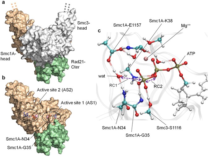
Figure 1

Model overview and description of the QM region. (a) Overview of the structural model of the complex formed by the human Smc1A-head (brown), Smc3-head (grey) and Rad21-Cter (green) domains. The dashed lines indicate the direction along which the un-modelled coiled coils would extend towards the hinge domain. (b) Location of active site 1 (AS1) and active site 2 (AS2). The Smc1A-head (brown) and Rad21-Cter (green) domains are shown, while the Smc3-head domain is not represented to reveal the location of the active sites. The location of the Smc1A-N34 and Smc1A-G35 residues is indicated. (c) QM region of AS1. The atoms in the QM region of the QM/MM MD simulations of AS1 are represented by coloured ball and sticks. The MM regions of the ATP (white ball and sticks) and protein backbone (transparent grey ribbons) are shown. The positions of the catalytic water (wat), residues Smc1A-N34, Smc1A-G35, Smc1A-K38, Smc1A-E1157 and Smc3-S1116, magnesium ion (Mg++) and ATP molecule are indicated. Reaction coordinates 1 (RC1) and 2 (RC2) are indicated by purple arrows.
