Figure 1

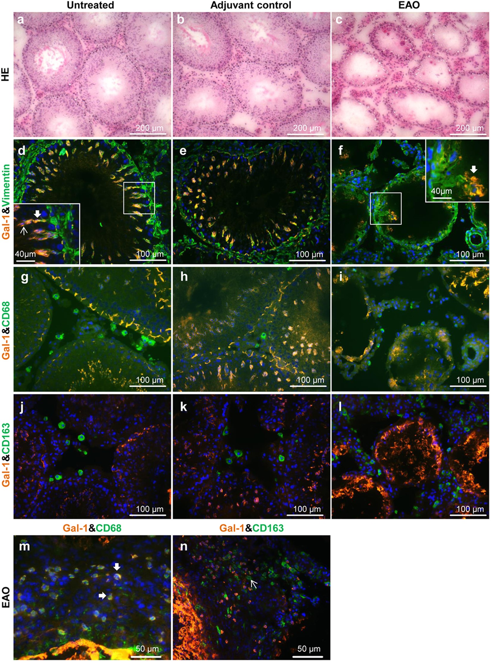
In normal rat testes Gal-1 is expressed mainly in Sertoli cells and germ cells but not in macrophages. Hematoxylin-eosin (HE) staining in cryostat sections from normal (a), adjuvant control (b) and EAO (c) rat testes. Localization of Gal-1 (Alexa 546, orange) in normal (d,g,j), adjuvant control (e,h,k) and EAO (f,i,l,m,n) testis. Vimentin (Alexa 488, green) was used as a marker of Sertoli cells (d,e,f). Insets show Gal-1 (Alexa 546, orange) stained in germ cells (thin arrow) and Sertoli cells (thick arrow) (d,f). Staining of Gal-1 and CD68 (Alexa 488, green) or CD163 (Alexa 488, green) in the region of granulomas (m,n). Testicular macrophages were stained with CD68 and CD163 antibodies. Gal-1 was expressed in some CD68 macrophages (m) found around granulomas (thick arrow), but not in CD163 macrophages (n) (thin arrow).
