Figure 1
From: Involvement of the habenula in the pathophysiology of autism spectrum disorder

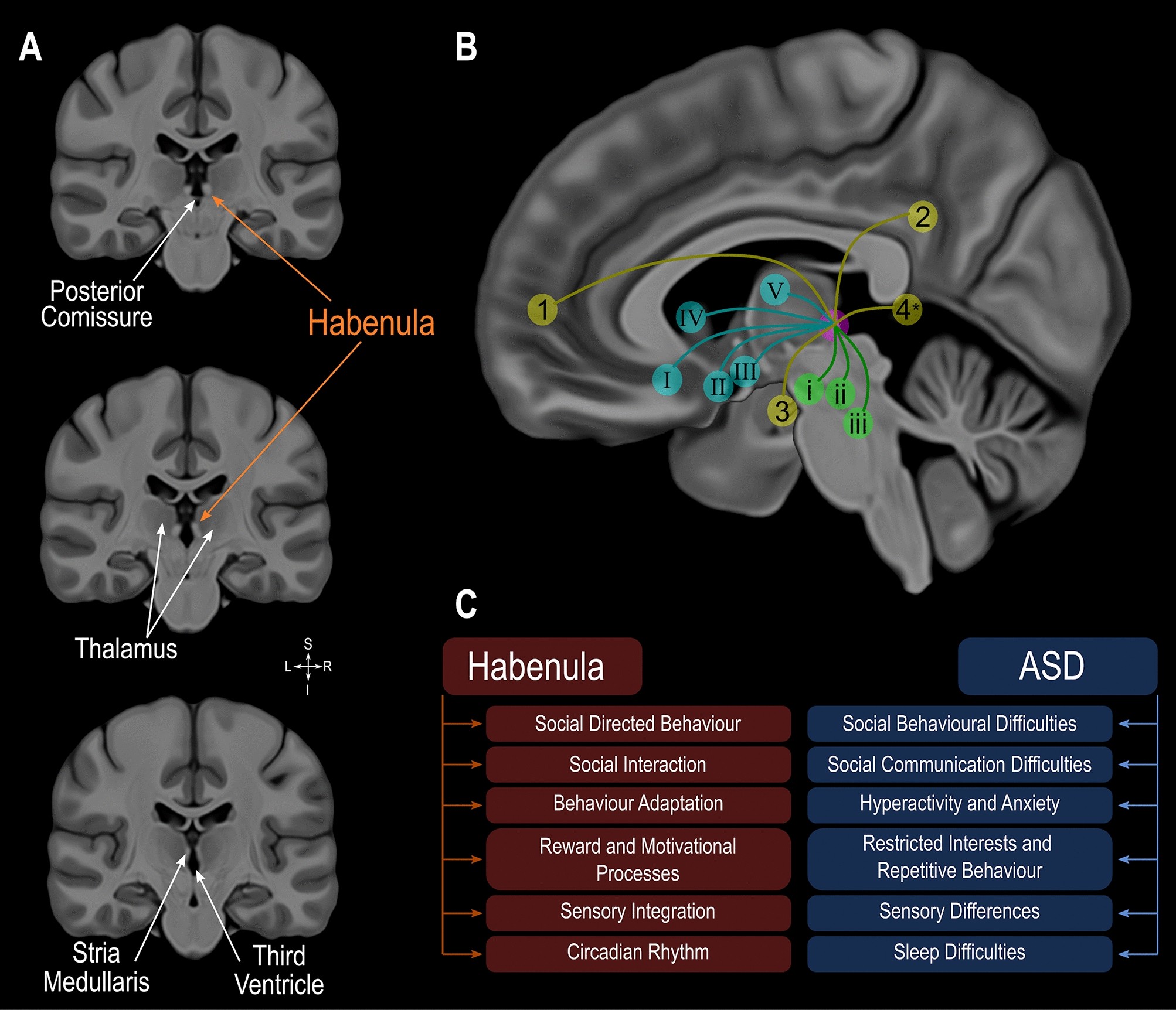
Habenula anatomy, boundaries and connections displayed using a high-resolution, high contrast template by Neudorfer and colleagues50. (A) Coronal slices illustrating the location of the Habenula, a structure appearing bright (hyperintense) on T1 weighted magnetic resonance images, surrounding structures and its boundaries. (B) Diagram illustrating the connectivity of the habenula. Cortical regions in yellow: (1) medial prefrontal cortex; (2) cingulate gyrus; (3) hippocampus and parahippocampal gyrus; (4) posterior insula (*estimated location). Subcortical regions in blue: (I) basal forebrain; (II) hypothalamus; (III) nucleus basalis of Meynert; IV, basal ganglia; V, thalamus. Brainstem regions in green: (i) ventral tegmental area; (ii) substantia nigra; (iii) periaqueductal grey—raphe nuclei. (C) Functions that the Habenula is critically involved in and differences found in autism spectrum disorder. ASD: autism spectrum disorder.
