Figure 1
From: Distribution and possible function of galanin about headache and immune system in the rat dura mater

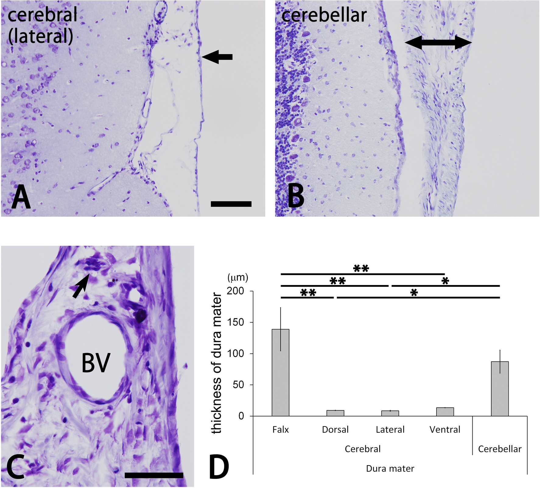
Photomicrographs for cresyl violet-stained cerebral and cerebellar dura mater (A–C). The lateral portion of the cerebral dura mater is thin (arrow in A), whereas the cerebellar dura mater is very thick (right and left arrows in B). The thick dura mater (C) contains a large blood vessel (BV) and nerve bundle (arrow). Bars = 100 µm (A) and 50 µm (C). Panels (A) and (B) are at the same magnification. Bar graphs indicate the thickness of the cerebral and cerebellar dura mater (D). The cerebral falx and cerebellar dura mater were significantly thicker than other portions of the cerebral dura mater (Tukey’s test *p < 0.05; **p < 0.01). Data were obtained from three sections of the cerebral falx; three to six sections of the dorsal, lateral and ventral portions of the cerebral dura mater; and two sections of the cerebellar dura mater in each of the four animals.
