Fig. 1
From: Direct visualization of human myosin II force generation using DNA origami-based thick filaments

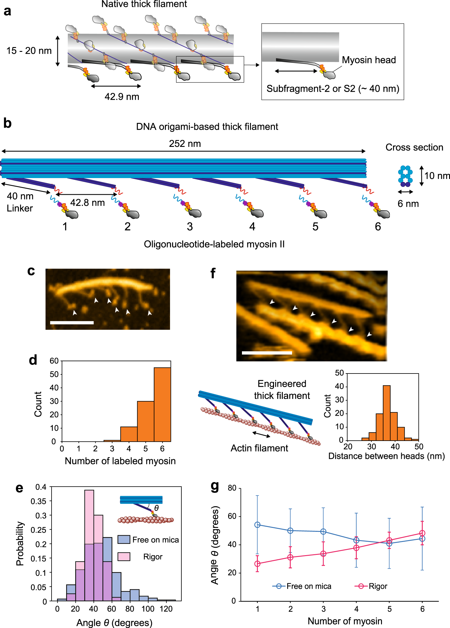
Design and AFM observation of DNA origami-based thick filaments. a Schematic of a native thick filament. Although myosin II in a thick filament forms a dimer, only the monomeric form is shown for simplicity. b Schematic of a DNA origami-based engineered thick filament. A 10-helix-bundle DNA origami rod structure (~10 nm x ~6 nm at the orthogonal cross section, blue) is the backbone of the filament, and six 2-helix-bundles of 40 nm length (“Linker”, dark blue) are separated along the backbone. 21-bp (~7 nm) oligonucleotide handles (red) are attached at the tip of the linkers for hybridization with complementary antihandles (light blue) labeled on myosin S1. c An AFM image of the DNA origami-based thick filament on mica. Arrowheads indicate myosin heads. Scale bar, 100 nm. d The number of myosin S1 labeled to a thick filament. Average occupancy, 5.4. e Histograms of angles (θ) between the linker and the backbone in the free state on mica and rigor state on lipid. Mean values were 47 ± 19o (SD) in the free state (n = 302) and 39 ± 9o (SD) in the rigor state (n = 80). f An AFM image of the actomyosin complex on lipid. Arrowheads indicate myosin heads. Cartoon of the complex and histogram of the distance between heads on actin (two-headed arrow) are also shown below. The mean value of the distances was 36 ± 3.5 nm (SD). Scale bar, 100 nm. g Average angles at each linker position in the free state on mica and rigor state on lipid. n = 47 for myosin 1 and 51 for myosins 2–6 in the free state. n = 11 for myosin 1, 18 for myosin 2 and 23 for myosins 3–6 in the rigor state. Data were obtained from at least 10 independent experiments. Error bars indicate SD.
