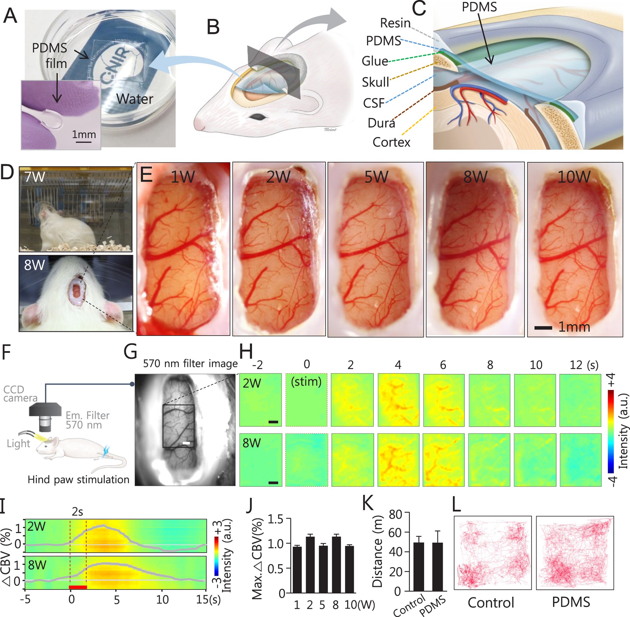
Figure 1: PDMS chronic cranial window development and installation and cortical vascular structural and functional observations during long-term studies.
From: A soft, transparent, freely accessible cranial window for chronic imaging and electrophysiology

(A) A hydrophobic and transparent square-type PDMS pad is floating on water and the clearly visible logo is placed under the culture dish. An insert image shows the bending feature of PDMS. (B,C) Schematics of the cranial window of rodents with flexible PDMS covering. (D) An animal with PDMS window behaves naturally in its home-cage 7 weeks post-implantation (top, and Supplementary Video S1) and no sign of inflammation around the implantation is visible 8 weeks post-implantation (bottom). (E) Magnified cortical images of the PDMS cranial window at 1, 2, 5, 8 and 10 weeks post-implantation. Despite temporal sequences clear vasculature is evident in all images. (F–J) Optical recording of intrinsic signal (ORIS) via PDMS window. (F) ORIS was performed with total haemoglobin weighted 570 nm wavelength during electrical stimulation (1 mA, 3 Hz, 2 s) of the left hind paw in three animals. (G) One example shows a clear brain surface image obtained using ORIS (scale bar: 1 mm). (H) A robust CBV change was observed after stimulation. The time course of peak intensity changes was obtained from the active region of 2.96 × 2.96 mm2 (Black box in G, scale bar: 1 mm). Group-averaged time course of CBV change at 8-weeks post-implantation (n = 3, mean ± SEM) is plotted in (I) and the average maximum CBV changes (mean ± SEM) at 1, 2, 5, 8 and 10 weeks post-implantation (n = 3 for each group) are shown in (J). Red bar in (I): 2 s stimulation period. In comparison with CBV changes at 1w, no significant differences were found in CBV changes at 2w (p = 0.70), 5w (p = 0.87), 8w (p = 0.67), and 10w (p = 0.79) with two-tailed Student’s t-test. (K,L) Home cage activity of a rat with a PDMS cranial window was studied and compared with control rat. (K) No significant moving distance difference was found for 30 minutes activity in each group (n = 3, p = 0.99, two-tailed Student’s t-test). The graph shows in mean ± SD. A single animal’s navigation pattern from each group is shown in (L).
