Volume 2
-
No. 12 December 2005
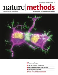
Cytoskeleton in differentiating neuroblastoma cells. Image courtesy of Torsten Wittmann, Department of Cell and Tissue Biology, University of California, San Francisco.
Focus
-
No. 11 November 2005
Carbohydrates on an array. Artistic adaptation by Erin Boyle. Review p817, Articles p845 and p851
-
No. 10 October 2005
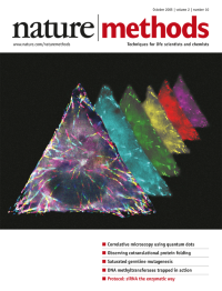
Pseudocolored timelapse images showing microtubule dynamics in a triangular eukaryotic cell patterned with an anisotropic solid microetching approach. Picture courtesy of Bartosz Grzybowski; artistic adaptation by Erin Boyle following suggestions of Bartosz Grzybowski and Kristiana Kandere-Grzybowska. Brief Communication p739
-
No. 9 September 2005
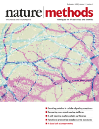
Confocal microscopic images of hippocampal neurons expressing PSD-95-GFP with calibrated fluorescent microspheres and stained with anti-MAP2 and anti-synapsin. Picture courtesy of Shigeo Okabe; artistic adaptation by Erin Boyle. Article p677
-
No. 8 August 2005
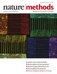
Rat neurons growing in a microfluidic culture platform. Neuritic processes extend from the neurons in the somal compartment through the microgrooves toward the axonal compartment. Picture courtesy of Noo Li Jeon; artistic adaptation by Erin Boyle. Article p 599
-
No. 7 July 2005
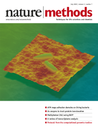
Atomic force microscopy topographic image of living mycobacteria on a polymer. Photograph courtesy of Yves F.Dufrêne; artistic adaptation by Erin Boyle. Article p515
-
No. 6 June 2005
Collage of images depicting immunohistochemistry stains in mouse brain sections. Photograph courtesy or Ari Waisman; artistic adaptation by Erin Boyle following suggestions by Saskia Hemmers. Article p 419
-
No. 5 May 2005
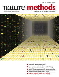
Image of DNA hybridized to a long oligonucleotide microarray. Photograph courtesy of Brenda Weis; artistic adaptation by Erin Boyle. Articles p337, 345 and 351
-
No. 4 April 2005
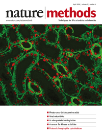
Immunofluorescence microscopy image of a mouse kidney section; lateral cell membranes are stained with an antibody against cadherin (green), nuclei with DAPI (red). Micrograph courtesy of Dario Neri; artistic adaptation by Erin Boyle. Article p291
-
No. 3 March 2005
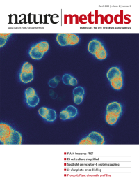
Time-lapse FRET images of a transmembrane receptor, tagged with FLAsH and a flourescent protein, after agonist stimulation. Pictures courtesy of Martin Lohse; artistic adaptation by Erin Boyle. Article p. 171.
-
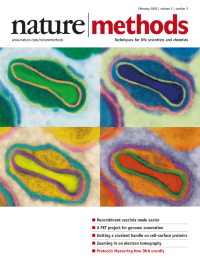
No. 2 February 2005
Transmission electron microscopy of a mature vaccinia virus particle in a thin section of an infected cell. Electron micrograph courtesy of Andrea Weisberg, Laboratory of Viral Diseases, NIAID, NIH; artistic adaptation by Erin Boyle.
-
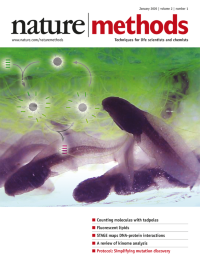
No. 1 January 2005
As tadpoles add diversity to a pond, molecular tadpoles, made up of a protein head and a DNA tail, will add important techniques to the molecular quantification toolbox. Photograph courtesy of Richard Nicholson, Christleton, UK; adaptation by Erin Boyle.