Volume 21
-
No. 12 December 2024
Method of the Year 2024: spatial proteomicsSpatial proteomics is our pick for Method of the Year 2024. The cover illustrates cellular neighborhoods within tumors, revealing the diverse and complex microenvironments that shape tumor biology.
See Editorial
-
No. 11 November 2024
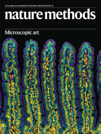
Microscopic artAn image of a section of small intestine from a mouse won fourth place in the Nikon Small World 2024 Photomicrography competition.
See Editorial
-
No. 10 October 2024
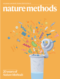
20 years of Nature MethodsThis month, Nature Methods celebrates its 20th anniversary with a special feature.
See Editorial
-
No. 9 September 2024
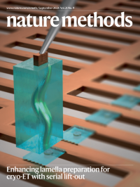
Enhancing lamella preparation for cryo-ET with serial lift-outArtistic representation of the sectioning step in a focused ion beam-based sample preparation technique, Serial Lift-Out. A block of vitreously frozen biological material (here, a C. elegans L1 larva embedded in buffer) is attached to a micromanipulator needle and transferred to a rectangular-mesh copper electron microscopy grid to be serially sectioned.
See Article
-
No. 8 August 2024
Focus on advanced AI in biologyAdvanced artificial intelligence (AI)-based methods are having a transformative impact on biological research, as explored in this special issue.
See Editorial
-
No. 7 July 2024
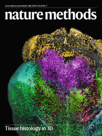
Tissue histology in 3Dc-Fos+ neuronal mapping of whole mouse brains using DELiVR reveals cancer-induced brain activity changes.
See Kaltenecker et al.
-
No. 6 June 2024
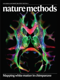
Mapping white matter in chimpanzeeTransverse view of a whole-brain tract-density reconstruction of white matter pathways in the chimpanzee brain. Color indicates tissue orientation and brightness encodes density of reconstructed fiber streamlines.
See Eichner et al.
-
No. 5 May 2024
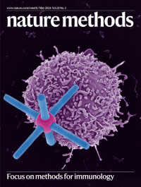
Focus on methods for immunologyA scanning electron microscope image captures the dynamic interplay between a CD19-hexapod biomimetic antigen-presenting structure and an anti-CD19 CAR-T cell.
See Huang et al.
-
No. 4 April 2024
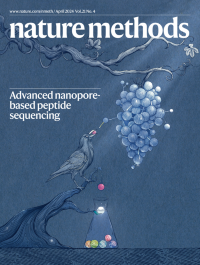
Advanced nanopore-based peptide sequencingPeptide sequencing by nanopore: a crow drops grapes into a pitcher with a narrow neck, representing the cleavage of a peptide into amino acids and their subsequent detection by a modified nanopore.
See Zhang et al.
-
No. 3 March 2024
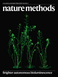
Brighter autonomous bioluminescenceAutonomously glowing Arabidopsis thaliana plants express an improved version of the fungal bioluminescence pathway.
See Shakhova et al.
-
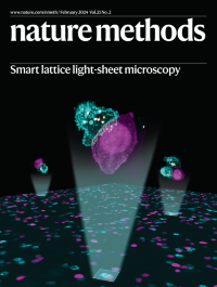
No. 2 February 2024
Smart lattice light-sheet microscopySmart lattice light-sheet microscopy captures rare cellular events. The image shows immune synapses formed between cytotoxic T lymphocytes (cyan) and tumor cells (magenta) within a population of cultured cells. Cytotoxic granules are shown in yellow.
See Shi et al.
-
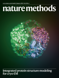
No. 1 January 2024
Integrated protein structure modeling for cryo-EMDeepMainmast builds structures of protein complexes from cryo-electron microscopy maps. It uses deep learning to identify key atom positions in the density, which are then connected to build fragment structures. Fragments are combined into a full structure, which is refined to atomic detail.
See Terashi et al.