Volume 4
-
No. 12 December 2007
Artist's rendition of the 'dual DNA' assay. Two DNA molecules (green and yellow) attached to beads (blue) are manipulated by optical tweezers (red cones) in a laminar flow cell with multiple buffer solutions running parallel to each other. Artistic rendering and image by Maarten Noom. Article p1031
-
No. 11 November 2007
Specific genomic enrichment methods, represented by Erin Boyle, based on an idea by Thomas Albert. The image of DNA fragments was drawn on a microarray by spotting 4 oligonucleotides according to the image pattern (the oligonucleotides were identical to the oligonucleotide later hybridized to the array, except for increasing number of mismatches, corresponding to different shades of grey in the black and white image) (courtesy of Thomas Albert, NimbleGen Systems). Brief Communications p903, 907, Article p931, News and Views p891
-
No. 10 October 2007
An artistic interpretation of mass spectrometrybased proteomics by Erin Boyle,using images from Nertila Siuti,Troy Hayes and Neil Kelleher.
-
No. 9 September 2007
Artistic interpretation by Erin Boyle of a Katushka fluorescent protein expression in a Xenopus laevis frog bearing the Katushka transgene under the control of a cardiac actin promoter. Protein structure representation is derived from the related protein egFP611. Frog image provided by Konstantin Lukyanov and Andrey Zaraisky.
-
No. 8 August 2007
Artistic interpretation of an enzyme activity assay using a macrocyclic receptor for a fluorescent dye. The macrocycle binds the enzyme substrate weakly but the product strongly, such that the product displaces the fluorescent dye, restoring its fluorescence. Cover by Erin Boyle.
-
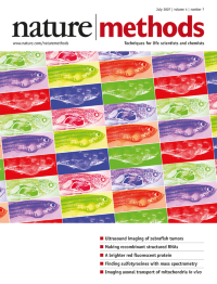
No. 7 July 2007
Artistic interpretation by Erin Boyle of zebrafish images provided by Wolfram Goessling and Trista North. Photographs, histological sections and ultrasound images all show a large abdominal tumor.
-
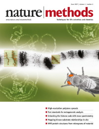
No. 6 June 2007
Artistic interpretation of high-resolution Drosophila polytene chromosome spreads by Erin Boyle, based on images provided by Dmitri Novikov. High-resolution light microscopic images were obtained by using precision devices to spread the chromosomes. Article p483
-
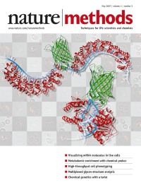
No. 5 May 2007
Artistic interpretation of mRNA visualization in living cells by Erin Boyle based on designs and images provided by Jeffrey Gerst, Natalia Broude and Yoshio Umezawa. Ribbon structures of the proteins were redrawn using Protein Data Bank (PDB) coordinates. Brief Communication p409, Articles p413 and p421.
-
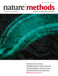
No. 4 April 2007
Fluorescence image of GFP-labeled neurons in a mouse hippocampus obtained by ultramicroscopy. Cover by Erin Boyle using an image by Hans-Ulrich Dodt. Article p331
-
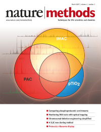
No. 3 March 2007
Artist's rendition of a comparative evaluation of three phosphoproteomics enrichment methods followed by mass spectrometry analysis, using images provided by Ruedi Aebersold and coworkers. Cover by Erin Boyle. Article p231
-
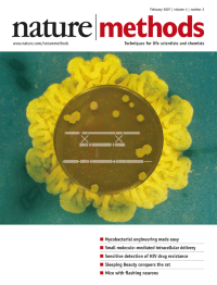
No. 2 February 2007
Recombineering in mycobacteria using mycobacteriophage-derived recombination proteins. Cover by Erin Boyle, using images from Graham Hatfull, Julia van Kessel and Tom Harper.
-
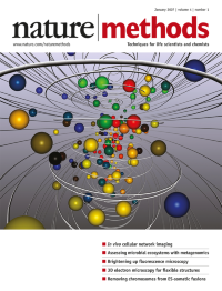
No. 1 January 2007
Artist's rendition of three-dimensional line-scan technology for two-photon microscopic imaging of in vivo cellular networks, based on images provided by Fritjof Helmchen. Cover by Erin Boyle. Article p73.