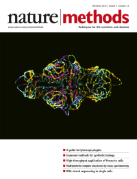
Volume 9
-
No. 12 December 2012
Single-molecule Förster resonance energy transfer (smFRET) and multiparameter fluorescence detection (2D histogram) help solve the structure of a protein-DNA complex using distance restraints derived from measured distances between fluorophore pairs (red and green spheres). Image courtesy of Thomas Peulen and Hugo Sanabria. Article p1218
-
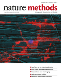
No. 11 November 2012
This photograph of the blood-brain barrier in a live zebrafish embryo took first place in the 2012 Nikon Small World photomicrography competition. The confocal microscope image (reprinted with permission from Nikon) was taken by Jennifer Peters and Michael Taylor of St. Jude Children's Research Hospital. Other images from this year's competition are on display at http://www.nikonsmallworld.com/.
-
No. 10 October 2012
Ultrafast force-clamp spectroscopy characterizes load dependence of the interaction between a single bead-attached myosin molecule (structure at center) and an actin filament (long polymer). Optical traps (red cones) apply constant forces on the actin ends. Artistic rendering by Marco Capitanio. Article p1013
-
No. 9 September 2012
An oligodendrocyte stained for myelin basic protein (red) and DNA (blue) interacts with polystyrene nanofibers. Cover by Matt Hansen, based on an image provided by Seonok Lee and Jonah R. Chan.
-
No. 8 August 2012
An Escherichia coli 'community' gene regulatory network, made by combining the predictions of several network inference methods tested in the DREAM5 challenge. Cover by Erin Dewalt, based on a design and image provided by Daniel Marbach and Gustavo Stolovitzky.
-
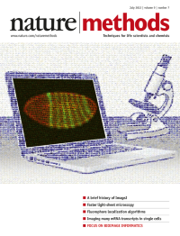
No. 7 July 2012
Mosaic image created with the CoverMaker plug-in in Fiji using images of Drosophila gene-expression patterns from histochemistry or fluorescence RNA in situ hybridization experiments. Image courtesy of Pavel Tomancak.
-
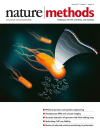
No. 6 June 2012
The cover illustration depicts the integration of foreign sequences into the DNA of Escherichia coli at targeted sites. Image provided by Jaehwan Jeong (concept) and Dong Su Jang (illustration). Brief Communication p591
-
No. 5 May 2012
The cover image is an artistic rendering of a Caenorhabditis elegans worm expressing a single-chain variable antibody fused to GFP that recognizes heparan sulfate glycans in the extracellular space. Image was provided by Hannes Bülow and Matthew Attreed. Cover design by Erin Dewalt. Brief Communication p477
-
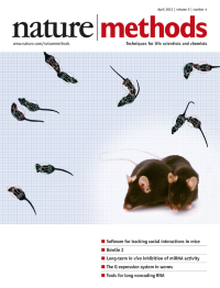
No. 4 April 2012
Physics models superimposed on video images of interacting mice, as tracked using the software MiceProfiler by Olivo-Marin and colleagues. Cover design by Erin Dewalt, based on images provided by Fabrice de Chaumont and Arnaud Cressant. Article p410
-
No. 3 March 2012
A simulated cell forms dynamic E-cadherin–mediated contact with a neighboring cell (the latter is not shown) over 60 minutes. The color code depicts relative concentrations of E-cadherin from lowest (blue) to highest (red). Morphological dynamics were simulated using a cellular Potts model; geometry and relative concentrations were exported from Simmune. Cover design by Erin Dewalt, based on images and simulations provided by Martin Meier-Schellersheim. Article p283
-
No. 2 February 2012
Image of an original oil painting by Emily Ferenczi–a graduate student in the laboratory of Karl Deisseroth–inspired by Wassily Kandinsky's Yellow-Red-Blue. Although individual elements are open to interpretation, this painting depicts an experiment involving optogenetic control of neuronal activity. Analysis p159
-
No. 1 January 2012
Genome editing with engineered endonucleases, our choice for Method of the Year 2011, allows scientists to make precise, targeted changes in the genome. Cover design by Erin Dewalt. Special feature starts on p23.