Volume 14
-
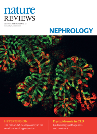
No. 12 December 2018
A mouse renal tubule expressing the mTmG reporter, which was grown ex vivo from primary cell organoids using a new 3D culture system for modelling pathogenesis in autosomal dominant polycystic kidney disease.
-
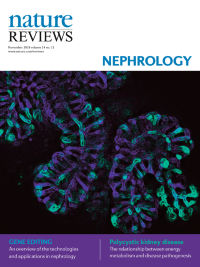
No. 11 November 2018
A mouse renal tubule expressing the mTmG reporter, which was grown ex vivo from primary cell organoids using a new 3D culture system for modelling pathogenesis in autosomal dominant polycystic kidney disease.
-
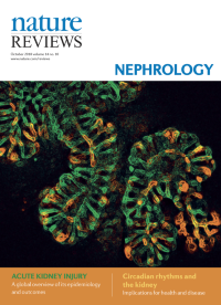
No. 10 October 2018
A mouse renal tubule expressing the mTmG reporter, which was grown ex vivo from primary cell organoids using a new 3D culture system for modelling pathogenesis in autosomal dominant polycystic kidney disease.
-
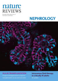
No. 9 September 2018
A mouse renal tubule expressing the mTmG reporter, which was grown ex vivo from primary cell organoids using a new 3D culture system for modelling pathogenesis in autosomal dominant polycystic kidney disease.
-
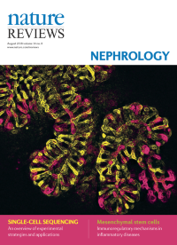
No. 8 August 2018
A mouse renal tubule expressing the mTmG reporter, which was grown ex vivo from primary cell organoids using a new 3D culture system for modelling pathogenesis in autosomal dominant polycystic kidney disease.
-
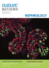
No. 7 July 2018
A mouse renal tubule expressing the mTmG reporter, which was grown ex vivo from primary cell organoids using a new 3D culture system for modelling pathogenesis in autosomal dominant polycystic kidney disease.
-
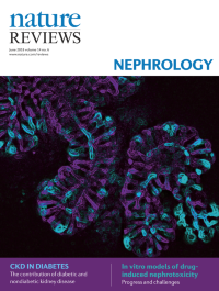
No. 6 June 2018
A mouse renal tubule expressing the mTmG reporter, which was grown ex vivo from primary cell organoids using a new 3D culture system for modelling pathogenesis in autosomal dominant polycystic kidney disease.
-
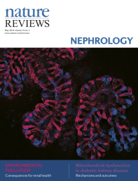
No. 5 May 2018
A mouse renal tubule expressing the mTmG reporter, which was grown ex vivo from primary cell organoids using a new 3D culture system for modelling pathogenesis in autosomal dominantpolycystic kidney disease. Cover image provided by Eryn E. Dixon of the Woodward Laboratory in the Department of Physiology and the Baltimore PKD Research and Clinical Core Center at the University of Maryland School of Medicine, Baltimore, MD, USA.
-
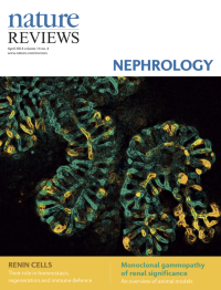
No. 4 April 2018
A mouse renal tubule expressing the mTmG reporter, which was grown ex vivo from primary cell organoids using a new 3D culture system for modelling pathogenesis in autosomal dominantpolycystic kidney disease. Cover image provided by Eryn E. Dixon of the Woodward Laboratory in the Department of Physiology and the Baltimore PKD Research and Clinical Core Center at the University of Maryland School of Medicine, Baltimore, MD, USA.
-
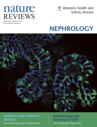
No. 3 March 2018
A mouse renal tubule expressing the mTmG reporter, which was grown ex vivo from primary cell organoids using a new 3D culture system for modelling pathogenesis in autosomal dominantpolycystic kidney disease. Cover image provided by Eryn E. Dixon of the Woodward Laboratory in the Department of Physiology and the Baltimore PKD Research and Clinical Core Center at the University of Maryland School of Medicine, Baltimore, MD, USA.
-
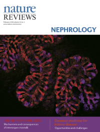
No. 2 February 2018
A mouse renal tubule expressing the mTmG reporter, which was grown ex vivo from primary cell organoids using a new 3D culture system for modelling pathogenesis in autosomal dominantpolycystic kidney disease. Cover image provided by Eryn E. Dixon of the Woodward Laboratory in the Department of Physiology and the Baltimore PKD Research and Clinical Core Center at the University of Maryland School of Medicine, Baltimore, MD, USA.
-
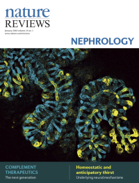
No. 1 January 2018
A mouse renal tubule expressing the mTmG reporter, which was grown ex vivo from primary cell organoids using a new 3D culture system for modelling pathogenesis in autosomal dominantpolycystic kidney disease. Cover image provided by Eryn E. Dixon of the Woodward Laboratory in the Department of Physiology and the Baltimore PKD Research and Clinical Core Center at the University of Maryland School of Medicine, Baltimore, MD, USA.