Volume 11
-
No. 12 December 2014
Cover image supplied by Arnulf Stenzl, Department of Urology, Eberhard-Karls-University Tuebingen, Tuebingen, Germany. Volume rendering is used for the reconstruction and 3D visualization of 2D images collected by CT or MRI. This image shows volume rendering of the abdominal aorta in a 73-year-old man who presented with severe flank pain and was subsequently diagnosed with renal atherosclerotic disease.
-
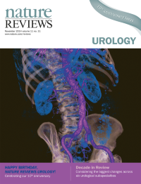
No. 11 November 2014
Cover image supplied by Arnulf Stenzl, Department of Urology, Eberhard-Karls-University Tuebingen, Tuebingen, Germany. Volume rendering is used for the reconstruction and 3D visualization of 2D images collected by CT or MRI. This image shows volume rendering of the abdominal aorta in a 73-year-old man who presented with severe flank pain and was subsequently diagnosed with renal atherosclerotic disease.
-
No. 10 October 2014
Cover image supplied by Arnulf Stenzl, Department of Urology, Eberhard-Karls-University Tuebingen, Tuebingen, Germany. Volume rendering is used for the reconstruction and 3D visualization of 2D images collected by CT or MRI. This image shows volume rendering of the abdominal aorta in a 73-year-old man who presented with severe flank pain and was subsequently diagnosed with renal atherosclerotic disease.
-
No. 9 September 2014
Cover image supplied by Arnulf Stenzl, Department of Urology, Eberhard-Karls-University Tuebingen, Tuebingen, Germany. Volume rendering is used for the reconstruction and 3D visualization of 2D images collected by CT or MRI. This image shows volume rendering of the abdominal aorta in a 73-year-old man who presented with severe flank pain and was subsequently diagnosed with renal atherosclerotic disease.
-
No. 8 August 2014
Cover image supplied by Arnulf Stenzl, Department of Urology, Eberhard-Karls-University Tuebingen, Tuebingen, Germany. Volume rendering is used for the reconstruction and 3D visualization of 2D images collected by CT or MRI. This image shows volume rendering of the abdominal aorta in a 73-year-old man who presented with severe flank pain and was subsequently diagnosed with renal atherosclerotic disease.
-
No. 7 August 2014
Cover image supplied by Arnulf Stenzl, Department of Urology, Eberhard-Karls-University Tuebingen, Tuebingen, Germany. Volume rendering is used for the reconstruction and 3D visualization of 2D images collected by CT or MRI. This image shows volume rendering of the abdominal aorta in a 73-year-old man who presented with severe flank pain and was subsequently diagnosed with renal atherosclerotic disease.
-
No. 6 June 2014
over image supplied by Arnulf Stenzl, Department of Urology, Eberhard-Karls-University Tuebingen, Tuebingen, Germany. Volume rendering is used for the reconstruction and 3D visualization of 2D images collected by CT or MRI. This image shows volume rendering of the abdominal aorta in a 73-year-old man who presented with severe flank pain and was subsequently diagnosed with renal atherosclerotic disease.
-
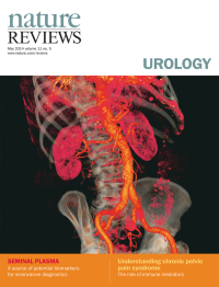
No. 5 May 2014
Cover image supplied by Arnulf Stenzl, Department of Urology, Eberhard-Karls-University Tuebingen, Tuebingen, Germany. Volume rendering is used for the reconstruction and 3D visualization of 2D images collected by CT or MRI. This image shows volume rendering of the abdominal aorta in a 73-year-old man who presented with severe flank pain and was subsequently diagnosed with renal atherosclerotic disease.
-
No. 4 April 2014
Cover image supplied by Arnulf Stenzl, Department of Urology, Eberhard-Karls-University Tuebingen, Tuebingen, Germany. Volume rendering is used for the reconstruction and 3D visualization of 2D images collected by CT or MRI. This image shows volume rendering of the abdominal aorta in a 73-year-old man who presented with severe flank pain and was subsequently diagnosed with renal atherosclerotic disease.
-
No. 3 March 2014
Cover image supplied by Arnulf Stenzl, Department of Urology, Eberhard-Karls-University Tuebingen, Tuebingen, Germany. Volume rendering is used for the reconstruction and 3D visualization of 2D images collected by CT or MRI. This image shows volume rendering of the abdominal aorta in a 73-year-old man who presented with severe flank pain and was subsequently diagnosed with renal atherosclerotic disease.
-
No. 2 February 2014
Cover image supplied by Arnulf Stenzl, Department of Urology, Eberhard-Karls-University Tuebingen, Tuebingen, Germany. Volume rendering is used for the reconstruction and 3D visualization of 2D images collected by CT or MRI. This image shows volume rendering of the abdominal aorta in a 73-year-old man who presented with severe flank pain and was subsequently diagnosed with renal atherosclerotic disease.
-
No. 1 January 2014
Cover image supplied by Arnulf Stenzl, Department of Urology, Eberhard-Karls-University Tuebingen, Tuebingen, Germany. Volume rendering is used for the reconstruction and 3D visualization of 2D images collected by CT or MRI. This image shows volume rendering of the abdominal aorta in a 73-year-old man who presented with severe flank pain and was subsequently diagnosed with renal atherosclerotic disease.