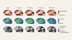
A brain reconstruction from an MRI scan using Oxford Brain Diagnostics’ software allows researchers to explore neurodegenerative changes.Credit: Oxford Brain Diagnostics
Oxford Brain Diagnostics is a spin-off from the University of Oxford, UK.
Neuroscientist Steven Chance spent 20 years looking at brain tissue through microscopes and examining magnetic resonance imaging (MRI) scans. During this time, he became increasingly interested in the information gap between the two. Histological analysis using microscopes reveals cellular scale changes, but only after death. MRI, by contrast, is safe for use in people, but can visualize only relatively large-scale changes. Chance wanted the best of both worlds: “an index of cellular scale changes, in life”, he says. “That’s the trajectory I was on.” Such changes reveal the neurodegeneration underlying numerous forms of dementia, including Alzheimer’s disease. There are still no therapies that alter these processes, and the World Health Organization estimates that 152 million people worldwide will be living with dementia by 2050.
Read more about The Spinoff Prize
In 2010, Chance teamed up with Mark Jenkinson, a neuroimaging researcher at the University of Oxford’s Wellcome Centre for Integrative Neuroimaging, UK. Eight years later, they founded Oxford Brain Diagnostics, with the aim of using specialized brain-imaging analysis to diagnose neurodegenerative conditions earlier, differentiate between them better and guide treatment decisions.
The company uses a form of MRI called diffusion tensor imaging, which measures dispersion of water through tissue. The cortex, the brain’s outermost region, is organized into cellular columns, giving an orderly microstructure to the tissue. The MRI system developed by Chance and Jenkinson measures disruption to that structure (see ‘Finding Alzheimer’s early’). “If there’s more gaps, the molecules pass through in different directions much more,” says Chance. “That starts to happen as you lose axons and cells.” Essentially, the researchers developed a non-invasive measure of brain-tissue integrity, enabling them to quantify neurodegeneration in people. “Once we had a set of measurements we felt were good at bridging this gap, it seemed natural to want to take that into the real world,” says Chance.

Initial funding of about £110,000 (US$136,000) enabled the company to demonstrate three important things, Chance says. First, using post-mortem brains, the team confirmed that its imaging metrics closely correspond to histological measures of cellular disruption that are sensitive to disease progression. Second, it analysed scans from a group of people with mild cognitive impairment. The MRI system could differentiate between people who progressed to Alzheimer’s within one year, two years or not at all. Finally, a collaboration with an Italian group validated the capability of the technique to differentiate between healthy older people and individuals with Alzheimer’s. “We had robustness in terms of the technology, and thresholds of distinction between the healthy and disease states, from two different scanners, in different parts of the world,” says Chance. “That was very encouraging.”
Part of Nature Outlook: The Spinoff Prize 2020
Although Alzheimer’s is a focus, it presents specific challenges for an approach based on brain changes. “What defines Alzheimer’s disease is the presence of amyloid and tau, which cannot be measured with MRI,” says neuroscientist Susan Landau of the University of California, Berkeley. Most researchers think Alzheimer’s is caused by a build-up of toxic forms of these two proteins. Indeed, studies using positron emission tomography (PET) scans suggest that amyloid might begin to accumulate 15–20 years before symptoms appear.
However, there is some controversy regarding whether amyloid is always the main cause of Alzheimer’s. Doubts are driven partly by the failure of drug trials to clear amyloid, and partly by the observation that amyloid burden does not correlate closely with Alzheimer’s symptoms. Measuring neurodegeneration directly sidesteps these issues. “Ultimately, we’re talking about the stuff you think with, and you don’t think with tau and amyloid,” says Chance. “By looking at the breakdown in the fine structure, we’re agnostic with respect to which protein caused which molecule to do what.”
Effect not cause
The MRI method that Oxford Brain Diagnostics is developing should prove more sensitive than approaches that measure changes in size of the hippocampus — the main structural biomarker used previously. “This type of imaging is really going to the physiological problems of the disease,” says neurobiologist George Perry of the University of Texas at San Antonio. “These are diseases of misconnected neurons.”
By focusing more on effect rather than cause, it means that the technique could work for a range of conditions. A study published last year, for instance, showed that the technology was sensitive to damage found in multiple sclerosis, a disease involving the breakdown of the myelin sheaths that insulate neural wiring1. The company also has results showing that the technique can differentiate between different ‘tauopathies’, including progressive supranuclear palsy, corticobasal degeneration and some forms of frontotemporal dementia (FTD), with accuracies of 90–95%. FTD is a common type of early onset dementia with subtypes that can be difficult to reliably diagnose, but the team has shown that its method can also distinguish between the three main FTD subtypes with 76% accuracy2. “With the specificity they have, what they report right now is already highly meaningful, because it could augment neuropsychological tests,” says Perry.
For neurodegenerative conditions, the main contenders in the disease biomarker market now are PET imaging using radiotracer ligands that bind to proteins, and analysis of cerebrospinal fluid. Blood-based measures are also under development. The company’s approach has some advantages. “It’s less invasive than cerebrospinal fluid and less expensive than amyloid or tau imaging,” says Perry.
But the start-up needs to demonstrate it has something to add. “How these measures compare with a clinician’s diagnosis, or cognitive tests, is really important,” says Landau. “Because it’s expensive, you have to show the scan gets you more than paper and pencil, or computerized tests, would,” says Landau.
Chance sees value in the diagnostic technology even in the absence of disease-modifying therapies. The company’s tests could help to differentiate between the “worried well”, and someone whose health might be about to deteriorate, he says. Ultimately, Chance wants people to view brain health the way they do heart health. “Everyone knows about blood pressure, what do you use for the brain?” he says. “This is missing.”
 The Spinoff Prize 2020
 44 firms highlighted in The Spinoff Prize 2020
 CageCapture: designing a molecule to filter out pollutants
 Caristo Diagnostics: taking a fresh look at CT scans
 EpiVario: mixing psychotherapy and small-molecule drugs
 EraCal Therapeutics: a new drug candidate for obesity
 Forkhead BioTherapeutics: developing a diabetes pill
 MiWEndo Solutions: using microwave technology to improve colonoscopies
 PredictImmune: a blood test to tailor treatment for bowel condition
 Scailyte: simplifying difficult diagnoses
 Sibel Health: designing vital-sign sensors for delicate skin
 Softsonics: a device to take way to blood-pressure readings continuously
 Temprian Therapeutics: developing a gene-based treatment for vitiligo