
Fig. 6: AIF2 KO has no effect on caspase-dependent and independent apoptosis.
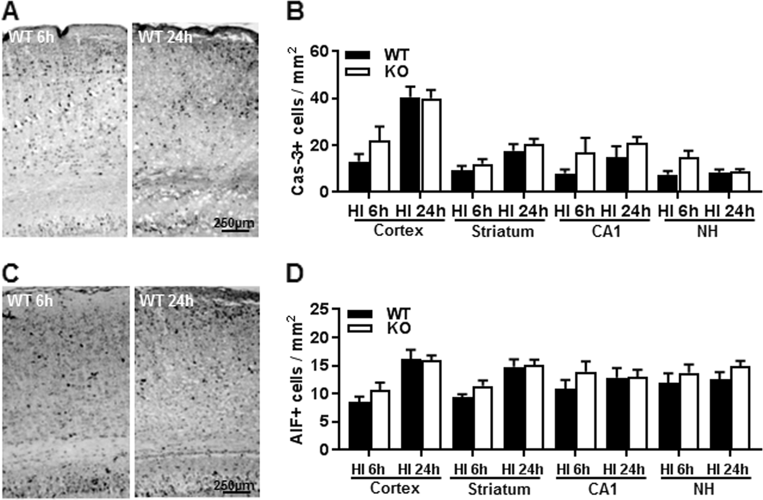
a Representative images show staining for the active form of caspase-3 at 6 and 24 h after HI. b Quantification of the number of active caspase-3-positive cells in WT and KO mice at 6 and 24 h post-HI. The bar graph has been divided into four sets for the cortex (CX), striatum (Str), CA1, and nuclei habenula (NH). There were no significant differences in the numbers of active caspase-3-positive cells. c Representative images showing the staining for the active form of AIF at 6 and 24 h after HI. d Quantification of the AIF-positive cells (nuclear AIF immunostaining) in WT and KO at 6 and 24 h post-HI. There were no significant differences between WT and KO at 6 h (n = 9/group) or 24 h (n = 8/group) after HI
