
Abstract
Since the outbreak of severe acute respiratory syndrome (SARS) 18 years ago, a large number of SARS-related coronaviruses (SARSr-CoVs) have been discovered in their natural reservoir host, bats1,2,3,4. Previous studies have shown that some bat SARSr-CoVs have the potential to infect humans5,6,7. Here we report the identification and characterization of a new coronavirus (2019-nCoV), which caused an epidemic of acute respiratory syndrome in humans in Wuhan, China. The epidemic, which started on 12 December 2019, had caused 2,794 laboratory-confirmed infections including 80 deaths by 26 January 2020. Full-length genome sequences were obtained from five patients at an early stage of the outbreak. The sequences are almost identical and share 79.6% sequence identity to SARS-CoV. Furthermore, we show that 2019-nCoV is 96% identical at the whole-genome level to a bat coronavirus. Pairwise protein sequence analysis of seven conserved non-structural proteins domains show that this virus belongs to the species of SARSr-CoV. In addition, 2019-nCoV virus isolated from the bronchoalveolar lavage fluid of a critically ill patient could be neutralized by sera from several patients. Notably, we confirmed that 2019-nCoV uses the same cell entry receptor—angiotensin converting enzyme II (ACE2)—as SARS-CoV.
Main
Coronaviruses have caused two large-scale pandemics in the past two decades, SARS and Middle East respiratory syndrome (MERS)8,9. It has generally been thought that SARSr-CoV—which is mainly found in bats—could cause a future disease outbreak10,11. Here we report on a series of cases caused by an unidentified pneumonia disease outbreak in Wuhan, Hubei province, central China. This disease outbreak—which started from a local seafood market—has grown substantially to infect 2,761 people in China, is associated with 80 deaths and has led to the infection of 33 people in 10 additional countries as of 26 January 202012. Typical clinical symptoms of these patients are fever, dry cough, breathing difficulties (dyspnoea), headache and pneumonia. Disease onset may result in progressive respiratory failure owing to alveolar damage (as observed by transverse chest computerized-tomography images) and even death. The disease was determined to be caused by virus-induced pneumonia by clinicians according to clinical symptoms and other criteria, including a rise in body temperature, decreases in the number of lymphocytes and white blood cells (although levels of the latter were sometimes normal), new pulmonary infiltrates on chest radiography and no obvious improvement after treatment with antibiotics for three days. It appears that most of the early cases had contact history with the original seafood market; however, the disease has now progressed to be transmitted by human-to-human contact.
Samples from seven patients with severe pneumonia (six of whom are sellers or deliverymen from the seafood market), who were admitted to the intensive care unit of Wuhan Jin Yin-Tan Hospital at the beginning of the outbreak, were sent to the laboratory at the Wuhan Institute of Virology (WIV) for the diagnosis of the causative pathogen (Extended Data Table 1). As a laboratory investigating CoV, we first used pan-CoV PCR primers to test these samples13, given that the outbreak occurred in winter and in a market—the same environment as SARS infections. We found five samples to be PCR-positive for CoVs. One sample (WIV04), collected from the bronchoalveolar lavage fluid (BALF), was analysed by metagenomics analysis using next-generation sequencing to identify potential aetiological agents. Of the 10,038,758 total reads—of which 1,582 total reads were retained after filtering of reads from the human genome—1,378 (87.1%) sequences matched the sequence of SARSr-CoV (Fig. 1a). By de novo assembly and targeted PCR, we obtained a 29,891-base-pair CoV genome that shared 79.6% sequence identity to SARS-CoV BJ01 (GenBank accession number AY278488.2). High genome coverage was obtained by remapping the total reads to this genome (Extended Data Fig. 1). This sequence has been submitted to GISAID (https://www.gisaid.org/) (accession number EPI_ISL_402124). Following the name given by the World Health Organization (WHO), we tentatively call it novel coronavirus 2019 (2019-nCoV). Four more full-length genome sequences of 2019-nCoV (WIV02, WIV05, WIV06 and WIV07) (GISAID accession numbers EPI_ISL_402127–402130) that were more than 99.9% identical to each other were subsequently obtained from four additional patients using next-generation sequencing and PCR (Extended Data Table 2).
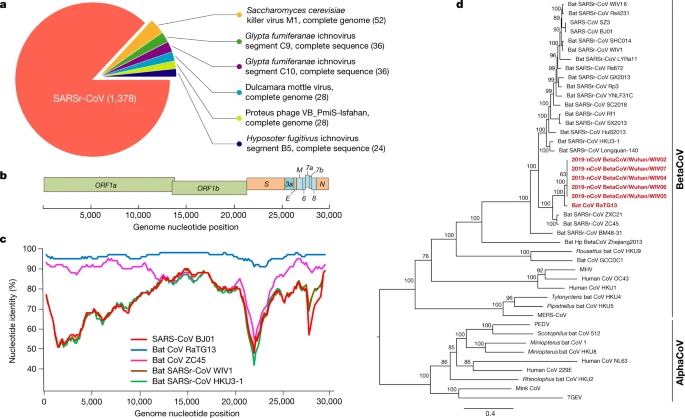
a, Metagenomics analysis of next-generation sequencing of BALF from patient ICU06. b, Genomic organization of 2019-nCoV WIV04. M, membrane. c, Similarity plot based on the full-length genome sequence of 2019-nCoV WIV04. Full-length genome sequences of SARS-CoV BJ01, bat SARSr-CoV WIV1, bat coronavirus RaTG13 and ZC45 were used as reference sequences. d, Phylogenetic tree based on nucleotide sequences of complete genomes of coronaviruses. MHV, murine hepatitis virus; PEDV, porcine epidemic diarrhoea virus; TGEV, porcine transmissible gastroenteritis virus.The scale bars represent 0.1 substitutions per nucleotide position. Descriptions of the settings and software that was used are included in the Methods.
The virus genome consists of six major open-reading frames (ORFs) that are common to coronaviruses and a number of other accessory genes (Fig. 1b). Further analysis indicates that some of the 2019-nCoV genes shared less than 80% nucleotide sequence identity to SARS-CoV. However, the amino acid sequences of the seven conserved replicase domains in ORF1ab that were used for CoV species classification were 94.4% identical between 2019-nCoV and SARS-CoV, suggesting that the two viruses belong to the same species, SARSr-CoV.
We then found that a short region of RNA-dependent RNA polymerase (RdRp) from a bat coronavirus (BatCoV RaTG13)—which was previously detected in Rhinolophus affinis from Yunnan province—showed high sequence identity to 2019-nCoV. We carried out full-length sequencing on this RNA sample (GISAID accession number EPI_ISL_402131). Simplot analysis showed that 2019-nCoV was highly similar throughout the genome to RaTG13 (Fig. 1c), with an overall genome sequence identity of 96.2%. Using the aligned genome sequences of 2019-nCoV, RaTG13, SARS-CoV and previously reported bat SARSr-CoVs, no evidence for recombination events was detected in the genome of 2019-nCoV. Phylogenetic analysis of the full-length genome and the gene sequences of RdRp and spike (S) showed that—for all sequences—RaTG13 is the closest relative of 2019-nCoV and they form a distinct lineage from other SARSr-CoVs (Fig. 1d and Extended Data Fig. 2). The receptor-binding spike protein encoded by the S gene was highly divergent from other CoVs (Extended Data Fig. 2), with less than 75% nucleotide sequence identity to all previously described SARSr-CoVs, except for a 93.1% nucleotide identity to RaTG13 (Extended Data Table 3). The S genes of 2019-nCoV and RaTG13 are longer than other SARSr-CoVs. The major differences in the sequence of the S gene of 2019-nCoV are the three short insertions in the N-terminal domain as well as changes in four out of five of the key residues in the receptor-binding motif compared with the sequence of SARS-CoV (Extended Data Fig. 3). Whether the insertions in the N-terminal domain of the S protein of 2019-nCoV confer sialic-acid-binding activity as it does in MERS-CoV needs to be further studied. The close phylogenetic relationship to RaTG13 provides evidence that 2019-nCoV may have originated in bats.
We rapidly developed a qPCR-based detection method on the basis of the sequence of the receptor-binding domain of the S gene, which was the most variable region of the genome (Fig. 1c). Our data show that the primers could differentiate 2019-nCoV from all other human coronaviruses including bat SARSr-CoV WIV1, which shares 95% identity with SARS-CoV (Extended Data Fig. 4a, b). Of the samples obtained from the seven patients, we found that six BALF and five oral swab samples were positive for 2019-nCoV during the first sampling, as assessed by qPCR and conventional PCR. However, we could no longer detect virus-positive samples in oral swabs, anal swabs and blood samples taken from these patients during the second sampling (Fig. 2a). However, we recommend that other qPCR targets, including the RdRp or envelope (E) genes are used for the routine detection of 2019-nCoV. On the basis of these findings, we propose that the disease could be transmitted by airborne transmission, although we cannot rule out other possible routes of transmission, as further investigation, including more patients, is required.
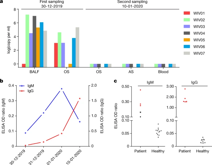
a, Molecular detection of 2019-nCoV in seven patients. Patient information can be found in Extended Data Tables 1, 2. Detection methods are described in the Methods. AS, anal swab; OS, oral swab. b, Dynamics of 2019-nCoV antibody levels in one patient who showed signs of disease on 23 December 2019 (ICU-06). OD ratio, optical density at 450–630 nm. The right and left y axes indicate ELISA OD ratios for IgM and IgG, respectively. c, Serological test of 2019-nCoV antibodies in five patients (Extended Data Table 2). The asterisk indicates data collected from patient ICU-06 on 10 January 2020. b, c, The cut-off was to 0.2 for the IgM analysis and to 0.3 for the IgG analysis, according to the levels of healthy controls.
For serological detection of 2019-nCoV, we used a previously developed nucleocapsid (N) protein from bat SARSr-CoV Rp3 as antigen for IgG and IgM enzyme-linked immunosorbent assays (ELISAs), as this protein shared 92% amino acid identity to N protein of 2019-nCoV (Extended Data Fig. 5) and showed no cross-reactivity against other human coronaviruses except SARSr-CoV7. We were only able to obtain five serum samples from the seven patients with viral infections. We monitored viral antibody levels in one patient (ICU-06) 7, 8, 9 and 18 days after the onset of disease (Extended Data Table 2). A clear trend was observed in the IgG and IgM titres, which increased over time, except that the IgM titre was decreased in the last sample (Fig. 2b). As a second analysis, we tested samples from 5 of the 7 virus-positive patients around 20 days after disease onset for the presence of viral antibodies (Extended Data Tables 1, 2). All patient samples—but not samples from healthy individuals—were strongly positive for viral IgG (Fig. 2b). There were also three IgM-positive samples, indicating an acute infection.
We next successfully isolated the virus (called 2019-nCoV BetaCoV/Wuhan/WIV04/2019) from both Vero E6 and Huh7 cells using the BALF sample of patient ICU-06. Clear cytopathogenic effects were observed in cells after incubation for three days (Extended Data Fig. 6a, b). The identity of the strain WIV04 was verified in Vero E6 cells by immunofluorescence microscopy using the cross-reactive viral N antibody (Extended Data Fig. 6c, d) and by metagenomics sequencing, most of the reads of which mapped to 2019-nCoV, and qPCR analysis showed that the viral load increased from day 1 to day 3 (Extended Data Fig. 6e, f). Viral particles in ultrathin sections of infected cells displayed a typical coronavirus morphology, as visualized by electron microscopy (Extended Data Fig. 6g). To further confirm the neutralization activity of the viral IgG-positive samples, we conducted serum-neutralization assays in Vero E6 cells using the five patient sera that were IgG-positive. We demonstrate that all samples were able to neutralize 100 TCID50 (50% tissue-culture-infective dose) of 2019-nCoV at a dilution of 1:40–1:80. We also show that this virus could be cross-neutralized by horse anti-SARS-CoV serum (gift from L.-F. Wang) at dilutions of 1:40; however, the potential for cross-reactivity with SARS-CoV antibodies needs to be confirmed with anti-SARS-CoV serum from humans (Extended Data Table 4).
ACE2 is known to be a cell receptor for SARS-CoV14. To determine whether 2019-nCoV also uses ACE2 as a cellular entry receptor, we conducted virus infectivity studies using HeLa cells that expressed or did not express ACE2 proteins from humans, Chinese horseshoe bats, civets, pigs and mice. We show that 2019-nCoV is able to use all ACE2 proteins, except for mouse ACE2, as an entry receptor to enter ACE2-expressing cells, but not cells that did not express ACE2, indicating that ACE2 is probably the cell receptor through which 2019-nCoV enters cells (Fig. 3). We also show that 2019-nCoV does not use other coronavirus receptors, such as aminopeptidase N (APN) and dipeptidyl peptidase 4 (DPP4) (Extended Data Fig. 7).
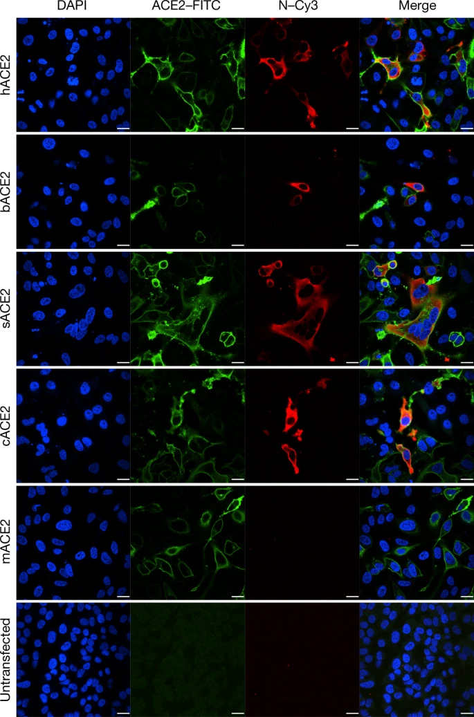
Determination of virus infectivity in HeLa cells that expressed or did not express (untransfected) ACE2. The expression of ACE2 plasmid with S tag was detected using mouse anti-S tag monoclonal antibody. hACE2, human ACE2; bACE2, ACE2 of Rhinolophus sinicus (bat); cACE2, civet ACE2; sACE2, swine ACE2 (pig); mACE2, mouse ACE2. Green, ACE2; red, viral protein (N); blue, DAPI (nuclei). Scale bars, 10 μm.
The study provides a detailed report on 2019-nCoV, the likely aetiological agent responsible for the ongoing epidemic of acute respiratory syndrome in China and other countries. Virus-specific nucleotide-positive and viral-protein seroconversion was observed in all patients tested and provides evidence of an association between the disease and the presence of this virus. However, there are still many urgent questions that remain to be answered. The association between 2019-nCoV and the disease has not been verified by animal experiments to fulfil the Koch’s postulates to establish a causative relationship between a microorganism and a disease. We do not yet know the transmission routine of this virus among hosts. It appears that the virus is becoming more transmissible between humans. We should closely monitor whether the virus continues to evolve to become more virulent. Owing to a shortage of specific treatments and considering the relatedness of 2019-nCoV to SARS-CoV, some drugs and pre-clinical vaccines against SARS-CoV could probably be used to treat this virus. Finally, considering the wide spread of SARSr-CoV in their natural reservoirs, future research should be focused on active surveillance of these viruses for broader geographical regions. In the long term, broad-spectrum antiviral drugs and vaccines should be prepared for emerging infectious diseases that are caused by this cluster of viruses in the future. Most importantly, strict regulations against the domestication and consumption of wildlife should be implemented.
Note added in proof: Since this paper was accepted, the ICTV has designated the virus as SARS-CoV-215; in addition, the WHO has released the official name of the disease caused by this virus, which is COVID-1916.
Methods
Data reporting
No statistical methods were used to predetermine sample size. The experiments were not randomized and the investigators were not blinded to allocation during experiments and outcome assessment.
Sample collection
Human samples, including oral swabs, anal swabs, blood and BALF samples were collected by Jinyintan hospital (Wuhan, China) with the consent of all patients and approved by the ethics committee of the designated hospital for emerging infectious diseases. Patients were sampled without gender or age preference unless indicated. For swabs, 1.5 ml DMEM containing 2% FBS was added to each tube. The supernatant was collected after centrifugation at 2,500 rpm, vortexing for 60 s and a standing period of 15–30 min. The supernatant from swabs or BALF (no pre-treatment) was added to either lysis buffer for RNA extraction or to viral transport medium for isolation of the virus. The viral transport medium was composed of Hank’s balanced salt solution (pH 7.4) containing BSA (1%), amphotericin (15 μg ml−1), penicillin G (100 units ml−1) and streptomycin (50 μg ml−1). Serum was separated by centrifugation at 3,000g for 15 min within 24 h of collection, followed by inactivation at 56 °C for 1 h, and was then stored at 4 °C until use.
Virus isolation, cell infection, electron microscopy and neutralization assay
The following cell lines were used for virus isolation in this study: Vero E6 and Huh7 cells, which were cultured in DMEM containing 10% FBS. All cell lines were tested and free of mycoplasma contamination, submitted for species identification and authenticated by morphological evaluation by microscopy. None of the cell lines was on the list of commonly misidentified cell lines (by ICLAC).
Cultured cell monolayers were maintained in their respective medium. The PCR-positive BALF sample from ICU-06 patient was spun at 8,000g for 15 min, filtered and diluted 1:2 with DMEM supplemented with 16 μg ml−1 trypsin before it was added to the cells. After incubation at 37 °C for 1 h, the inoculum was removed and replaced with fresh culture medium containing antibiotics (see below) and 16 μg ml−1 trypsin. The cells were incubated at 37 °C and observed daily for cytopathogenic effects. The culture supernatant was examined for the presence of virus by qRT–PCR methods developed in this study, and cells were examined by immunofluorescence microscopy using the anti-SARSr-CoV Rp3 N antibody that was generated in-house (1:1,000). Penicillin (100 units ml−1) and streptomycin (15 μg ml−1) were included in all tissue culture media.
Vero E6 cells were infected with the new virus at a multiplicity of infection (MOI) of 0.5 and collected 48 h after infection. Cells were fixed with 2.5% (w/v) glutaraldehyde and 1% osmium tetroxide, dehydrated through a graded series of ethanol concentrations (from 30 to 100%) and embedded with epoxy resin. Ultrathin sections (80 nm) of embedded cells were prepared, deposited onto Formvar-coated copper grids (200 mesh), stained with uranyl acetate and lead citrate, and analysed using a 200-kV Tecnai G2 electron microscope.
The virus neutralization test was carried out in a 96-well plate. The patient serum samples were heat-inactivated by incubation at 56 °C for 1 h before use. The serum samples were diluted to 1:10, 1:20, 1:40 or 1:80, and then an equal volume of virus stock was added and incubated at 37 °C for 60 min in a 5% CO2 incubator. Diluted horse anti-SARS-CoV serum or serum samples from healthy individuals were used as control. After incubation, 100 μl mixtures were inoculated onto a monolayer of Vero E6 cells in a 96-well plate for 1 h. Each serum was assessed in triplicate. After removing the supernatant, the plate was washed twice with DMEM medium. Cells were incubated with DMEM supplemented with 2% FBS for 3 days. Subsequently, the cells were checked for cytopathogenic effects.
RNA extraction and PCR
Whenever commercial kits were used, the manufacturer’s instructions were followed without modification. RNA was extracted from 200 μl of samples with the High Pure Viral RNA kit (Roche). RNA was eluted in 50 μl of elution buffer and used as the template for RT–PCR.
For qPCR analysis, primers based on the S gene of 2019-nCoV were designed: RBD-qF1, 5′-CAATGGTTTAACAGGCACAGG-3′; RBD-qR1, 5′-CTCAAGTGTCTGTGGATCACG-3′. RNA extracted as described above was used for qPCR using the HiScript II One Step qRT–PCR SYBR Green Kit (Vazyme Biotech). Conventional PCRs were also performed using the following primer pairs: ND-CoVs-951F, 5′-TGTKAGRTTYCCTAAYATTAC-3′; ND-CoVs-1805R, 5′-ACATCYTGATANARAACAGC-3′. The 20-μl qPCR reaction mix contained 10 μl 2× One Step SYBR Green mix, 1 μl One Step SYBR Green Enzyme mix, 0.4 μl 50× ROX Reference Dye 1, 0.4 μl of each primer (10 μM) and 2 μl template RNA. Amplification was performed as follows: 50 °C for 3 min, 95 °C for 30 s followed by 40 cycles consisting of 95 °C for 10 s and 60 °C for 30 s, and a default melting curve step in an ABI 7500 Real-time PCR machine.
Serological test
In-house anti-SARSr-CoV IgG and IgM ELISA kits were developed using SARSr-CoV Rp3 N protein as antigen, which shared more than 90% amino acid identity to all SARSr-CoVs2. For IgG analyses, MaxiSorp Nunc-immuno 96-well ELISA plates were coated (100 ng per well) overnight with recombinant N protein. Human sera were used at a dilution of 1:20 for 1 h at 37 °C. An anti-human IgG HRP-conjugated monoclonal antibody (Kyab Biotech) was used at a dilution of 1:40,000. The OD value (450–630 nm) was calculated. For IgM analyses, MaxiSorp Nunc-immuno 96-well ELISA plates were coated (500 ng per well) overnight with anti-human IgM (μ chain). Human sera were used at a 1:100 dilution for 40 min at 37 °C, followed by incubation with an anti-Rp3 N HRP-conjugated antibody (Kyab Biotech) at a dilution of 1:4,000. The OD value (450–630 nm) was calculated.
Examination of ACE2 receptor for 2019-nCoV infection
HeLa cells transiently expressing ACE2 were prepared using Lipofectamine 3000 (Thermo Fisher Scientific) in a 96-well plate; mock-transfected cells were used as controls. 2019-nCoV grown in Vero E6 cells was used for infection at a MOI of 0.5. APN and DPP4 were analysed in the same way. The inoculum was removed after absorption for 1 h and washed twice with PBS and supplemented with medium. At 24 h after infection, cells were washed with PBS and fixed with 4% formaldehyde in PBS (pH 7.4) for 20 min at room temperature. ACE2 expression was detected using a mouse anti-S tag monoclonal antibody and a FITC-labelled goat anti-mouse IgG H&L (Abcam, ab96879). Viral replication was detected using a rabbit antibody against the Rp3 N protein (generated in-house, 1:1,000) and a Cy3-conjugated goat anti-rabbit IgG (1:200, Abcam, ab6939). Nuclei were stained with DAPI (Beyotime). Staining patterns were examined using confocal microscopy on a FV1200 microscope (Olympus).
High-throughput sequencing, pathogen screening and genome assembly
Samples from patient BALF or from the supernatant of virus cultures were used for RNA extraction and next-generation sequencing (NGS) using BGI MGISEQ2000 and Illumina MiSeq 3000 sequencers. Metagenomic analysis was carried out mainly based on the bioinformatics platform MGmapper (PE_2.24 and SE_2.24). The raw NGS reads were first processed by Cutadapt (v.1.18) with minimum read length of 30 base pairs. BWA (v.0.7.12-r1039) was used to align reads to a local database with a filter hits parameter of 0.8 FMM ((match + mismatch)/read length ≥ fraction] value and minimum alignment score of 30. Parameters for post-processing of assigned reads were set to a minimum size normalized abundance of 0.01, minimum read count of 20 and were otherwise set to default parameters. A local nucleic acid database for human and mammals was used to filter reads of host genomes before mapping reads to the virus database. The results of the metagenomic analysis were displayed as pie charts using Microsoft Office 2010. NGS reads were assembled into genomes using Geneious (v.11.0.3) and MEGAHIT (v.1.2.9). PCR and Sanger sequencing was performed to fill gaps in the genome. 5′-rapid amplification of cDNA ends (RACE) was performed to determine the 5′-end of the genomes using a SMARTer RACE 5′/3′ kit (Takara). Genomes were annotated using the Clone Manager Professional Suite 8 (Sci-Ed Software).
Phylogenetic analysis
Routine sequence management and analysis was carried out using DNAStar. The sequence alignment of complete genome sequences was performed using MAFFT (v.7.307) with default parameters. The codon alignments of full-length S and RdRp gene sequences were converted from the corresponding protein alignments by PAL2NAL (v.14); the protein alignments were created by Clustal Omega (v.1.2.4) using default parameters. Maximum likelihood phylogenetic trees were generated using RAxML (v.0.9.0) with GTR+G substitution model and 1,000 bootstrap replicates.
Reporting summary
Further information on research design is available in the Nature Research Reporting Summary linked to this paper.
Data availability
Sequence data that support the findings of this study have been deposited in GISAID (https://www.gisaid.org/) with accession numbers EPI_ISL_402124, EPI_ISL_402127–EPI_ISL_402130 and EPI_ISL_402131; GenBank with accession numbers MN996527–MN996532; National Genomics Data Center, Beijing Institute of Genomics, Chinese Academy of Sciences (https://bigd.big.ac.cn/databases?lang=en) with accession numbers SAMC133236–SAMC133240 and SAMC133252.
Change history
28 September 2020
This article was amended to correct the Peer review information.
References
Li, W. et al. Bats are natural reservoirs of SARS-like coronaviruses. Science 310, 676–679 (2005).
Ge, X.-Y. et al. Isolation and characterization of a bat SARS-like coronavirus that uses the ACE2 receptor. Nature 503, 535–538 (2013).
Yang, L. et al. Novel SARS-like betacoronaviruses in bats, China, 2011. Emerg. Infect. Dis. 19, 989–991 (2013).
Hu, B. et al. Discovery of a rich gene pool of bat SARS-related coronaviruses provides new insights into the origin of SARS coronavirus. PLoS Pathog. 13, e1006698 (2017).
Menachery, V. D. et al. A SARS-like cluster of circulating bat coronaviruses shows potential for human emergence. Nat. Med. 21, 1508–1513 (2015).
Menachery, V. D. et al. SARS-like WIV1-CoV poised for human emergence. Proc. Natl Acad. Sci. USA 113, 3048–3053 (2016).
Wang, N. et al. Serological evidence of bat SARS-related coronavirus infection in humans, China. Virol. Sin. 33, 104–107 (2018).
Drosten, C. et al. Identification of a novel coronavirus in patients with severe acute respiratory syndrome. N. Engl. J. Med. 348, 1967–1976 (2003).
Zaki, A. M., van Boheemen, S., Bestebroer, T. M., Osterhaus, A. D. M. E. & Fouchier, R. A. M. Isolation of a novel coronavirus from a man with pneumonia in Saudi Arabia. N. Engl. J. Med. 367, 1814–1820 (2012).
Cui, J., Li, F. & Shi, Z. L. Origin and evolution of pathogenic coronaviruses. Nat. Rev. Microbiol. 17, 181–192 (2019).
Fan, Y., Zhao, K., Shi, Z.-L. & Zhou, P. Bat coronaviruses in China. Viruses 11, 210 (2019).
Wuhan Municipal Health Commission. Press statement related to novel coronavirus infection (in Chinese) http://wjw.wuhan.gov.cn/front/web/showDetail/2020012709194 (2020).
Poon, L. L. et al. Identification of a novel coronavirus in bats. J. Virol. 79, 2001–2009 (2005).
Li, W. et al. Angiotensin-converting enzyme 2 is a functional receptor for the SARS coronavirus. Nature 426, 450–454 (2003).
Gorbalenya, A. E. et al. Severe acute respiratory syndrome-related coronavirus — the species and its viruses, a statement of the Coronavirus Study Group. Preprint at https://www.biorxiv.org/content/10.1101/2020.02.07.937862v1 (2020).
WHO. WHO Director-General’s remarks at the media briefing on 2019-nCoV on 11 February 2020. https://www.who.int/dg/speeches/detail/who-director-general-s-remarks-at-the-media-briefing-on-2019-ncov-on-11-february-2020 (WHO, 11 February 2020).
Acknowledgements
We thank P. Zhang and A. Du from the WIV core facility centre for their help with producing transmission electron microscopy micrographs; H.-Z. Liu and P. Yu from WIV for bioinformatics analysis. This work was jointly supported by the Strategic Priority Research Program of the Chinese Academy of Sciences (CAS) (XDB29010101 to Z.-L.S. and XDB29010104 to P.Z.), China Natural Science Foundation for excellent scholars (81822028 to P.Z., 31770175 to Z.-L.S. and 31800142 to B.H.), Mega-Project for Infectious Disease from Minister of Science and Technology of the People’s Republic of China (2018ZX10305409-004-001 to P.Z.), Youth innovation promotion association of CAS (2019328 to X.-L.Y.).
Author information
Authors and Affiliations
Contributions
Z.-L.S., P.Z., Y.-Y.W. and G.-F.X. conceived the study. X.-G.W., C.-L.H., H.-D.C., F.D., Q.-J.C., F.-X.Z. and L.-L.L. collected patient samples. X.-L.Y., B.Y., W.Z., B.L., J.C., X.-S.Z., Y.L., H.G., R.-D.J., M.-Q.L., Y.C., X.W., X.-R.S. and K.Z. performed qPCR, serology and virus culturing experiments. L.Z., Y.Z., H.-R.S. and B.H. performed genome sequencing and annotations.
Corresponding author
Ethics declarations
Competing interests
The authors declare no competing interests.
Additional information
Peer review information Nature thanks the anonymous reviewers for their contribution to the peer review of this work.
Publisher’s note Springer Nature remains neutral with regard to jurisdictional claims in published maps and institutional affiliations.
Extended data figures and tables
Extended Data Fig. 1 NGS raw reads of sample WIV04 mapping to the 2019-nCoV sequence.
The x axis indicates the genome nucleotide position and the y axis represents the read depth of the mapping.
Extended Data Fig. 2 Phylogenetic trees based on the complete S and RdRp gene sequences of coronaviruses.
a, b, Phylogenetic trees on the basis of the gene sequences of S (a) and RdRp (b) are shown. 2019-nCoV and bat CoV RaTG13 are shown in bold and in red. The trees were constructed using the maximum likelihood method using the GTR + G substitution model with bootstrap values determined by 1,000 replicates. Bootstraps values of more than 50% are shown.
Extended Data Fig. 3 Amino acid sequence alignment of the S1 protein of the 2019-nCoV to SARS-CoV and selected bat SARSr-CoVs.
The receptor-binding motif of SARS-CoV and the homologous region of other coronaviruses are indicated by the red box. The key amino acid residues involved in the interaction with human ACE2 are numbered at the top of the aligned sequences. The short insertions in the N-terminal domain of the 2019-nCoV are indicated by the blue boxes. Bat CoV RaTG13 was obtained from R. affinis, found in Yunnan province. Bat CoV ZC45 was obtained from R. sinicus, found in Zhejiang province.
Extended Data Fig. 4 Molecular detection method used to detect 2019-nCoV.
a, Standard curve for qPCR primers. The PCR product of the S gene that was serial diluted in the range of 108 to 101 (lines from left to right) was used as a template. Primer sequences and experimental conditions are described in the Methods. b, Specificity of the qPCR primers. Nucleotide samples from the indicated pathogens were used.
Extended Data Fig. 5 Amino acid sequence alignment of the nucleocapsid protein of 2019-nCoV to bat SARSr-CoV Rp3 and SARS-CoV BJ01.
Bat SARSr-CoV Rp3 was obtained from R. sinicus, which is found in Guangxi province.
Extended Data Fig. 6 Isolation and antigenic characterization of 2019-nCoV.
a, b, Vero E6 cells are shown at 24 h after infection with mock virus (a) or 2019-nCoV (b). c, d, Mock-virus-infected (c) or 2019-nCoV-infected (d) samples were stained with rabbit serum raised against recombinant SARSr-CoV Rp3 N protein (red) and DAPI (blue). The experiment was conducted twice independently with similar results. e, The ratio of the number of reads related to 2019-nCoV among the total number of virus-related reads in metagenomics analysis of supernatants from Vero E6 cell cultures. f, Virus growth in Vero E6 cells. g, Viral particles in the ultrathin sections were imaged using electron microscopy at 200 kV. The sample was from virus-infected Vero E6 cells. The inset shows the viral particles in an intra-cytosolic vacuole.
Extended Data Fig. 7 Analysis of 2019-nCoV receptor usage.
Determination of virus infectivity in HeLa cells with or without the expression of human APN and DPP4. The expression of ACE2, APN and DPP4 plasmids with S tag were detected using mouse anti-S tag monoclonal antibody. ACE2, APN and DPP4 proteins (green), viral protein (red) and nuclei (blue) are shown. Scale bars, 10 μm.
Supplementary information
Rights and permissions
Open Access This article is licensed under a Creative Commons Attribution 4.0 International License, which permits use, sharing, adaptation, distribution and reproduction in any medium or format, as long as you give appropriate credit to the original author(s) and the source, provide a link to the Creative Commons license, and indicate if changes were made. The images or other third party material in this article are included in the article’s Creative Commons license, unless indicated otherwise in a credit line to the material. If material is not included in the article’s Creative Commons license and your intended use is not permitted by statutory regulation or exceeds the permitted use, you will need to obtain permission directly from the copyright holder. To view a copy of this license, visit http://creativecommons.org/licenses/by/4.0/.
About this article

Cite this article
Zhou, P., Yang, XL., Wang, XG. et al. A pneumonia outbreak associated with a new coronavirus of probable bat origin. Nature 579, 270–273 (2020). https://doi.org/10.1038/s41586-020-2012-7
Received:
Accepted:
Published:
Issue date:
DOI: https://doi.org/10.1038/s41586-020-2012-7
This article is cited by
-
Factors influencing the implementation of interventions for symptoms of posttraumatic stress disorder among hospital-based nurses and physicians during the COVID-19 pandemic: a scoping review
BMC Health Services Research (2025)
-
Brazilian attitudes towards Covid-19 and its relationship with personal conditions, political identification, empathy and susceptibility to accept fake news: a study during the third wave
Discover Public Health (2025)
-
Host taxonomy and environment shapes insectivore viromes and viral spillover risks in Southwestern China
Microbiome (2025)
-
Association between SARS-CoV-2 and stroke: perspectives from a metaumbrella-review
BMC Neurology (2025)
-
The role of artificial intelligence in pandemic responses: from epidemiological modeling to vaccine development
Molecular Biomedicine (2025)

Parvaiz
Nitazoxanide as a possible agent for the 2019-Novel Corona Virus infection
Isolation of the virus and testing of antiviral drugs is critical to the management of the cases at present as this outbreak seems to have shaped clearly worse than the SARS and the MERS-coV outbreaks reported in the earlier parts of this century. This would allow studying the antiviral efficacy of various antiviral agents against the virus. Pertinently combinations of antiviral agents can be tried. Additionally the antiparasitic agent nitazoxanide might be useful and may be worthwhile testing in patients who are sick based on the premise that the combination of nitazoxanide and oseltamivir was found to be superior to oseltamivir alone in influenza patients in a previous study. Nitazoxanide is a safe antiparasitic agent, widely prescribed and has been demonstrated to have antiviral properties which could be exploited in the current settings to treat severe cases in combination with investigational antiviral agents as also test its efficacy in the laboratory against cultured virus isolates.
Michael Siedel
just a few questions? 22 pages seven test persons only one had the virus detectable! all slight fever! 96 percent match! the rest of the other ingredients thrown together wildly? who cross-checks which laboratory level 4? why 9 days until approval by Nature, since submission when it is urgent? and then another 4 to upload 22 pages? Thank you very much! yes always the bats and always the markets and always test environment Wuhan! Dankeschön ihr Hausmeister Krause/ #fafnör
Michael Siedel member MinD Germany number 21165
lindsey rim Replied to Michael Siedel
i dont know what your talking about, but read this article plz if you know nothing about the coronacirus https://www.ladieshabits.co...
BiologyGeek
Tens of thousands of people are infected and hundreds died. We need to better understand how the bat coronavirus got transmitted to human patients in this 2020 outbreak. Many blame a wildlife market in Wuhan - but there are 661 cities in China and many of them, especially 41 in Guangdong where residents are known to like bushmeat, have such markets. Why didn't the outbreak start from any of the other 660 cities, especially the 41 in Guangdong - but why Wuhan? For example, the 2002-2003 SARS outbreak started from Fushan, Guangdong, but not Wuhan.
Moreover, according to Figure 1 of China CDC's recent report: https://www.nejm.org/doi/fu..., the first two patients had no link to the Wuhan wildlife market at all.
Now, if you look at this: there are 661 cities in China, but there is only one city, Wuhan, hosting a biosafety level 4 (BSL-4, or P4) lab. It's the only lab in China that captures bats and is authorized to study the bat coronavirus. The authors, as part of this BSL-4 lab, started to do so since 2015. Statistically, the bat coronavirus of the Wuhan outbreak is more likely to be from the only Wuhan BSL-4 lab that studies bat coronavirus in China, as opposed to be from the wildlife markets - Wuhan's wildlife market is among hundreds of wildlife markets existing in hundreds of cities in China. Is there any possibility that the bat coronavirus got leaked from this BSL-4 lab unintentionally through improper handling of samples and lab animals? This needs to be investigated.
BiologyGeek Replied to BiologyGeek
Also, we need to note that, as recently publicly pointed out by Dr. Yi Rao, President of Capital University of Medical University in China, Mrs. Yan-yi Wang - the Lab Director of the Wuhan BSL-4 lab as well as the supervisor of Dr. Zheng-li Shi - is professionally not qualified. News is here: https://www.backchina.com/n....
Moreover, the concern about the safety issue of the Wuhan BSL-4 lab was discussed as early as in 2017: https://www.nature.com/news...
The authors of this article are from the questioned Wuhan BSL-4 lab. The safety concern is serious. The possibility of an unintentional bat coronavirus leak causing this 2020 coronavirus outbreak should be at least discussed and investigated.
BiologyGeek Replied to BiologyGeek
An update on the questions on the origin of the coronavirus:
A recent news reported that China has denied the lab link to coronavirus. https://www.washingtontimes... "However, a Chinese government-funded study published Jan. 24 in the medical journal The Lancet by 29 Chinese scientists found that 13 of 41 victims surveyed had no connection to the wild animal market. Significantly, the first patient identified with the coronavirus was a man who reported pneumonia-like symptoms on Dec. 1 but had no connection to the market."
The mentioned Lancet paper is here: https://www.thelancet.com/j...
Who is the first patient? What happened to him? How come he had no link to the market? Why the coronavirus he contracted with "showed high sequence identity" to the "BatCoV RaTG13" stored in the lab of the authors?
Jason
some key data and logical connection is missing in order for the author to claim "We then found a short RdRp region from a bat coronavirus termed BatCoV RaTG13 which we previously detected in Rhinolophus affinis from Yunnan Province showed high sequence identity to nCoV-2019."
Duhnkuhn Hussein
Zheng-Li Shi will end up killing more people that Mao Zedong. That is impressive.
ben Replied to Duhnkuhn Hussein
How so?
bioconductor Sun
Where can the scientist world-wide get the raw NGS reads to review and re-analyze the squencing data?
madmaxNY
Wow, unbelievable that this passed editorial and peer review at Nature! Something very strange that they would rush a paper out that is even not seriously edited, with tons of grammatical errors, so how many scientific errors either intentional or otherwise? Reviewers, please identify yourselves and release your reviews, the extent to which you went to validate the results, whether you had access to the raw sequence data, etc. And as cases continue to be made that the senior author on this paper, Zheng-Li Shi, has spent her career engineering this type of virus, publishing papers on the ability to do this and so on:
https://www.zerohedge.com/g...
I guess the editors at Nature did not feel it necessary to question the integrity of these results and force a more in depth review and replication from others.
Sung-Joon Park
We performed a metagenomic approach with WHU RNA-seq data;
https://www.nature.com/arti...,
(https://openlooper.hgc.jp/s...).
Of note, WHU01 (SRR10903402) RNA-seq reads included statistically significant "Mycoplasma"-related reads, and the most probable species was "Mycoplasma hominis", while "Streptococcus"-related reads are probably not the human pathogen (contamination?) (https://openlooper.hgc.jp/f....
Although this paper has used virus genomes, we have prepared the subsets of the RNA-seq reads that could not be mapped to the human genome and over 325,000 RefSeq microbial genomes (inc. SARS).
Over 80% of the subset reads (R1 only) could be mapped to the nCoV genome (NC_045512), which may be greatly useful to characterize nCoV specific genome.
BatCoV
https://uploads.disquscdn.c... https://uploads.disquscdn.c... https://uploads.disquscdn.c... https://uploads.disquscdn.c...
Jodie Replied to BatCoV
Can we pm re these?
Rossana
The RdRp of RaTG13 has 100 % identity with the sequence BtCoV/4991
(KP876546) identified by Ge and colleagues in a Rhinolophus affinis bat in the
Yunnan province in 2013 as RaTG13. https://doi.org/10.1007/s12...
BtCoV/4991 is a novel beta-CoV, clearly separated from all known alpha- and beta-CoVs at that time. BtCoV/4991 differentiates from other bat CoVs also in the phylogenetic analysis carried out by Wang and colleagues https://doi.org/10.3390/v11...
Chen and colleagues identified BtCoV/4991 as the closest sequence to SARS-CoV-2 because RaTG13 had not yet been published at that time doi:
10.1080/22221751.2020.1725399.
How do BtCoV/4991 and RaTG13 relate to each other?
Kenneth McDonald
If anyone tries to post the now famous article "Manufactured Pandemic" on Facebook, they are threatened by Facebook, and a link to Flora Teoh's article "PCR tests for COVID-19 are specific for the novel coronavirus SARS-CoV-2 and do not detect other coronaviruses, contrary to claims in viral article and video" is forced onto the post as a supposed refutation.
Central to her claim the quick fix PCR test is accurate is her claim quote "Thanks to the efforts of scientists, we now know the full genome of SARS-CoV-2 [1]" and the link to "prove" this goes here. Can anyone here drag and drop a reply to me to show the "proof" of that from this article with a quote from it please?
If they span out in a centrifuge the SARS-CoV-2 virus, and mapped its RNA. it helps prove the credibility of the PCR Tests for SARS-CoV-2 detection, but not viral load. But if they really did isolate the virus that way, why not then put it in a healthy person and cause the disease and fulfill either the Koch's or the River's postulates? That Dr Andrew Kaufman says have not been fulfilled, so therefore calling SARS-CoV-2 an infectious disease is outside of accepted science. So when did you fulfil the River's or Koch's postulates? (several questions there).
Kenneth McDonald
Why is it that only one scientist, Flora Teoh, steps forward and says the PCR Test used on the first 3 million SARS-CoV-2 infections and death rate statistics is accurate, and shows viral load? Why don't hundreds step forward and put their reputations on the line? If Flora Teoh has this wrong, then the stats used to propose lockdowns in the Third World are bogus and unreliable, yet those lockdowns are said to cause an extra 20 million deaths of innocent women and children from starvation, as well as ruining the world economy so much we can barely help the millions of others who would usually be in danger of starvation. Thus it is MEGA important these PCR Tests are accurate, but we get ONE scientist stepping forward to tell us they are. Why?
Flora Teoh..... do you realise what you have done if you have this wrong???? MILLIONS OF CHILDREN AND BABIES may unnecessarily die in agony!
In my opinion she uses a reprehensible and clearly false tactic. She accuses the writer of the "Manufactured Pandemic" article of attacking the brilliant science of Kary Mullis of PCR in general. That is nonsense. I do not believe that for one second. The person obviously just thinks the SARS-CoV-2 Test method of use is wrong, especially if Kaufman is right that they compare the RNA they extract to an equally corrupted SARS-CoV-1 RNA strand, and it's only 80% similar.
Kenneth McDonald
I just wrote this to Health Feedback.org
QUOTE: "Covid-19 death predictions were wildly exaggerated by Imperial College. This helped to unreasonably form the idea lockdowns should occur in the Third World that will cause tens of millions of innocent women, children and babies to starve to death, in vastly higher numbers than the elderly might be saved to live a bit longer.
I agree the article "Manufactured Pandemic" did not have enough supporting evidence in it to prove some of its claims. All that proves is the article was badly written, not that the PCR Test did not at first have inefficient primers, or other possible failures. Your "refutation article" is being used to block anyone wanting to prove that they CAN come up with evidence missing from the Manufactured Pandemic article, as if you disproved the test has flaws, when all you proved was the article never had proof links.
Similarly Flora Teoh made the accusation the writer of the article was attacking Kary Mullis brilliant PCR technology, when all he/she was attacking was its particular use in this test. You then went on to say the person was "therefore" denying PCR can ever show viral load in OTHER tests or when he/she never. The ISSUE was did that specific test show viral load? As f it never we all have perhaps 10 viruses in our bodies in low viral load that do us no harm, (as Dr Andrew Kaufman points out) thus without viral load indication IN THAT ACTUAL TEST the test proves next to NOTHING.
You then allow no comments.
Do you realise that if you are saying the first 1 - 3 million stats are accurate when they are not that those stats (I believe are bogus) are being used to starve to death TENS OF MILLIONS of children in the Third World? AND your work is being used to stifle freedom of speech as well? Then you allow no comments to refute you?
Please reply to this message."
prajwol
I could not find accession numbers of reference sequence data that were used in phylogenetic analysis. If I were to replicate this study, how am I supposed to do so without any metadata table of the GenBank accession numbers of those reference sequences? How can Nature journal ignore these important component?
Samuel Soroaster
How could this ever get published in Nature? Did the reviewers not note that the most important reference namely the RATTG13 Sequence happend to be published by the same lab only a few days before publication? Why would they wait 7 years only until after the outbrake in Wuhan to publish the RATTG13 sequence? This needs to be followed up by the editor. https://www.ncbi.nlm.nih.go...
Monali C. Rahalkar
We have published a critical view on this paper in our preprint: https://www.preprints.org/m...
Alberto Maria Cattaneo
I also realized some inconsistency about the information available for RaTG13, have a look at my preprint on Researchgate: https://doi.org/10.13140/RG....
Zhou et al. mention to have collected RaTG13 samples from bats of the Yunnan province on 2013, but checking on GISAID, and all the other databases, RaTG13 was deposited only on 2020, no evidences that it was really sampled 7 years ago are available, apart "saying so".
Plus, in my preprint, I sequence-aligned the S-proteins of Covid-19 and RaTG13 with other Sars-Covs´, including Pangolin´s (Zhang et al. 2020 https://doi.org/10.1016/j.c... and I did not validated the same identity of the binding motif of the RBD that Zhang describes, neither I found evidences of the existence of a putative furin recognition motif in the S-proteins of Pangolin´s viruses.
All together, this rise further doubts about the origins of RaTG13. Supplementary evidences about the Sars-CoV-2 origins from Zhou et al. would be really appreciated.
Acron Naji
According to Shi's response to Science magazine, and her TV interview before, she doesn't have the isolated RaTG13 virus and any samples at her lab in Wuhan. This means there is no way to requecence RaTG13 scientifically and verify it existed or not in nature. Since it's crucial for the seeking of origin of SARS-Cov-2, I personally call for her retracting of this article.
https://www.sciencemag.org/...
Eric
Based on the fact it was submitted on January 20, 2020 and subsequently published on February 3, 2020, I could not fathom how it had been peer-reviewed so quickly. Upon further examination, I discovered that it has not been peer-reviewed at all. Per the publisher's website: "All peer-reviewed content will carry an anonymous statement of peer reviewer acknowledgement, and for those reviewers who give their consent, we will publish their names alongside the published article." No such statement exists for this article. I find this to be very odd, considering that this research has more than 2,300 citations and helped lay the foundation for the entire world community to declare an emergency of such remarkably unprecedented proportions.
https://www.nature.com/natu...
https://www.nytimes.com/201...
By comparison, see the "Peer review information" of a random article I selected in the Nature scientific journal about HIV research: "Nature thanks Nicolas Chomont, Philippe Lemey and the other, anonymous, reviewer(s) for their contribution to the peer review of this work."
https://www.nature.com/arti...
Top Cat ✓ᵛᵉʳᶦᶠᶦᵉᵈ Replied to Eric
It's late, so this is for future reference: Had you read that which you linked to, you could have fathomed how peer review could be done so quickly and not had to appeal to ignorance...
"Authors are usually informed within a week if the paper is not being considered. Most referees honour their prior agreement with Nature to deliver a report within seven days or other agreed time limit, and send their reports online."