Abstract
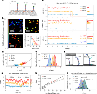
The intricate interplay between DNA and proteins is key for biological functions such as DNA replication, transcription and repair. Dynamic nanoscale observations of DNA structural features are necessary for understanding these interactions. Here we introduce graphene energy transfer with vertical nucleic acids (GETvNA), a method to investigate DNA–protein interactions that exploits the vertical orientation adopted by double-stranded DNA on graphene. This approach enables the dynamic study of DNA conformational changes via energy transfer from a probe dye to graphene, achieving spatial resolution down to the Ångström scale at subsecond temporal resolution. We measured DNA bending induced by adenine tracts, bulges, abasic sites and the binding of endonuclease IV. In addition, we observed the translocation of the O6-alkylguanine DNA alkyltransferase on DNA, reaching single base-pair resolution and detecting preferential binding to adenine tracts. This method promises widespread use for dynamical studies of nucleic acids and nucleic acid–protein interactions with resolution so far reserved for traditional structural biology techniques.
This is a preview of subscription content, access via your institution
Access options
Access Nature and 54 other Nature Portfolio journals
Get Nature+, our best-value online-access subscription
$32.99 / 30 days
cancel any time
Subscribe to this journal
Receive 12 print issues and online access
$259.00 per year
only $21.58 per issue
Buy this article
- Purchase on SpringerLink
- Instant access to the full article PDF.
USD 39.95
Prices may be subject to local taxes which are calculated during checkout


Data availability
Raw and processed data supporting the findings of this work are available on Zenodo at https://doi.org/10.5281/zenodo.13794574 (ref. 89).
Code availability
A home-made Python script used to process the raw data is available as Supplementary Software alongside example data. The Python scripts used to generate the plots presented in the figures of this work, based on processed data, are available on Zenodo at https://doi.org/10.5281/zenodo.13794574 (ref. 89).
References
Lerner, E. et al. Toward dynamic structural biology: two decades of single-molecule Förster resonance energy transfer. Science 359, eaan1133 (2018).
Ha, T. Single-molecule fluorescence methods for the study of nucleic acids. Curr. Opin. Struct. Biol. 11, 287–292 (2001).
Schuler, B. & Eaton, W. A. Protein folding studied by single-molecule FRET. Curr. Opin. Struct. Biol. 18, 16–26 (2008).
Lerner, E. et al. FRET-based dynamic structural biology: challenges, perspectives and an appeal for open-science practices. ife 10, e60416 (2021).
Quast, R. B. & Margeat, E. Single-molecule FRET on its way to structural biology in live cells. Nat. Methods 18, 344–345 (2021).
Wolff, J. O. et al. MINFLUX dissects the unimpeded walking of kinesin-1. Science 379, 1004–1010 (2023).
Deguchi, T. et al. Direct observation of motor protein stepping in living cells using MINFLUX. Science 379, 1010–1015 (2023).
Reinhardt, S. C. M. et al. Ångström-resolution fluorescence microscopy. Nature 617, 711–716 (2023).
Masullo, L. A., Lopez, L. F. & Stefani, F. D. A common framework for single-molecule localization using sequential structured illumination. Biophys. Rep. 2, 100036 (2022).
Sahl, S. J. et al. Direct optical measurement of intramolecular distances with angstrom precision. Science 386, 180–187 (2024).
Cole, F. et al. Super-resolved FRET and co-tracking in pMINFLUX. Nat. Photon 18, 478–484 (2024).
Kaminska, I. et al. Distance dependence of single-molecule energy transfer to graphene measured with DNA origami nanopositioners. Nano Lett. 19, 4257–4262 (2019).
Kamińska, I. et al. Graphene energy transfer for single-molecule biophysics, biosensing, and super-resolution microscopy. Adv. Mater. 33, 2101099 (2021).
Ghosh, A. et al. Graphene-based metal-induced energy transfer for sub-nanometre optical localization. Nat. Photonics 13, 860–865 (2019).
Weber, M. et al. MINSTED nanoscopy enters the Ångström localization range. Nat. Biotechnol. 41, 569–576 (2023).
Gwosch, K. C. et al. MINFLUX nanoscopy delivers 3D multicolor nanometer resolution in cells. Nat. Methods 17, 217–224 (2020).
Thevathasan, J. V. et al. Nuclear pores as versatile reference standards for quantitative superresolution microscopy. Nat. Methods 16, 1045–1053 (2019).
Secundo, F. Conformational changes of enzymes upon immobilisation. Chem. Soc. Rev. 42, 6250–6261 (2013).
Wolf, L. K., Gao, Y. & Georgiadis, R. M. Sequence-dependent DNA immobilization: specific versus nonspecific contributions. Langmuir 20, 3357–3361 (2004).
Hamlin, R. E., Dayton, T. L., Johnson, L. E. & Johal, M. S. A QCM study of the immobilization of β-galactosidase on polyelectrolyte surfaces: effect of the terminal polyion on enzymatic surface activity. Langmuir 23, 4432–4437 (2007).
Krause, S. et al. Graphene-on-glass preparation and cleaning methods characterized by single-molecule DNA origami fluorescent probes and raman spectroscopy. ACS Nano 15, 6430–6438 (2021).
Zähringer, J. et al. Combining pMINFLUX, graphene energy transfer and DNA-PAINT for nanometer precise 3D super-resolution microscopy. Light. Sci. Appl. 12, 70 (2023).
Minetti, C. A. S. A., Remeta, D. P., Dickstein, R. & Breslauer, K. J. Energetic signatures of single base bulges: thermodynamic consequences and biological implications. Nucleic Acids Res. 38, 97–116 (2010).
Joshua-Tor, L. et al. Three-dimensional structures of bulge-containing DNA fragments. J. Mol. Biol. 225, 397–431 (1992).
Koo, H.-S., Wu, H.-M. & Crothers, D. M. DNA bending at adenine · thymine tracts. Nature 320, 501–506 (1986).
Tolstorukov, M. Y., Virnik, K. M., Adhya, S. & Zhurkin, V. B. A-tract clusters may facilitate DNA packaging in bacterial nucleoid. Nucleic Acids Res. 33, 3907–3918 (2005).
Haran, T. E. & Mohanty, U. The unique structure of A-tracts and intrinsic DNA bending. Q. Rev. Biophys. 42, 41–81 (2009).
Rohs, R. et al. The role of DNA shape in protein–DNA recognition. Nature 461, 1248–1253 (2009).
Levin, J. D., Johnson, A. W. & Demple, B. Homogeneous Escherichia coli endonuclease IV. Characterization of an enzyme that recognizes oxidative damage in DNA. J. Biol. Chem. 263, 8066–8071 (1988).
Lorenz, M. et al. DNA bending induced by high mobility group proteins studied by fluorescence resonance energy transfer. Biochemistry 38, 12150–12158 (1999).
Hillisch, A., Lorenz, M. & Diekmann, S. Recent advances in FRET: distance determination in protein–DNA complexes. Curr. Opin. Struct. Biol. 11, 201–207 (2001).
Arnott, S. & Hukins, D. W. L. Optimised parameters for A-DNA and B-DNA. Biochem. Biophys. Res. Commun. 47, 1504–1509 (1972).
Lilley, D. M. Kinking of DNA and RNA by base bulges. Proc. Natl Acad. Sci. USA 92, 7140–7142 (1995).
Gu, H., Yang, W. & Seeman, N. C. DNA scissors device used to measure muts binding to DNA mis-pairs. J. Am. Chem. Soc. 132, 4352–4357 (2010).
Woźniak, A. K., Schröder, G. F., Grubmüller, H., Seidel, C. A. M. & Oesterhelt, F. Single-molecule FRET measures bends and kinks in DNA. Proc. Natl Acad. Sci. USA 105, 18337–18342 (2008).
Dornberger, U., Hillisch, A., Gollmick, F. A., Fritzsche, H. & Diekmann, S. Solution structure of a five-adenine bulge loop within a DNA duplex. Biochemistry 38, 12860–12868 (1999).
Shi, X., Beauchamp, K. A., Harbury, P. B. & Herschlag, D. From a structural average to the conformational ensemble of a DNA bulge. Proc. Natl Acad. Sci. USA 111, E1473–E1480 (2014).
Schreck, J. S., Ouldridge, T. E., Romano, F., Louis, A. A. & Doye, J. P. K. Characterizing the bending and flexibility induced by bulges in DNA duplexes. J. Chem. Phys. 142, 165101 (2015).
MacDonald, D., Herbert, K., Zhang, X., Polgruto, T. & Lu, P. Solution structure of an A-tract DNA bend. J. Mol. Biol. 306, 1081–1098 (2001).
Koo, H. S. & Crothers, D. M. Calibration of DNA curvature and a unified description of sequence-directed bending. Proc. Natl Acad. Sci. USA 85, 1763–1767 (1988).
Garcin, E. D. et al. DNA apurinic-apyrimidinic site binding and excision by endonuclease IV. Nat. Struct. Mol. Biol. 15, 515–522 (2008).
Hosfield, D. J., Guan, Y., Haas, B. J., Cunningham, R. P. & Tainer, J. A. Structure of the DNA repair enzyme endonuclease IV and Its DNA complex: double-nucleotide flipping at abasic sites and three-metal-ion catalysis. Cell 98, 397–408 (1999).
Stivers, J. T. 2-Aminopurine fluorescence studies of base stacking interactions at abasic sites in DNA: metal-ion and base sequence effects. Nucleic Acids Res. 26, 3837–3844 (1998).
Cuniasse, P. H., Fazakerley, G. V., Guschlbauer, W., Kaplan, B. E. & Sowers, L. C. The abasic site as a challenge to DNA polymerase: a nuclear magnetic resonance study of G, C and T opposite a model abasic site. J. Mol. Biol. 213, 303–314 (1990).
Chen, J., Dupradeau, F.-Y., Case, D. A., Turner, C. J. & Stubbe, J. Nuclear magnetic resonance structural studies and molecular modeling of duplex DNA containing normal and 4′-oxidized abasic sites. Biochemistry 46, 3096–3107 (2007).
Hoitsma, N. M. et al. AP-endonuclease 1 sculpts DNA through an anchoring tyrosine residue on the DNA intercalating loop. Nucleic Acids Res. 48, 7345–7355 (2020).
Mol, C. D., Izumi, T., Mitra, S. & Tainer, J. A. DNA-bound structures and mutants reveal abasic DNA binding by APE1 DNA repair and coordination. Nature 403, 451–456 (2000).
Zhu, C. et al. Tautomerization-dependent recognition and excision of oxidation damage in base-excision DNA repair. Proc. Natl Acad. Sci. 113, 7792–7797 (2016).
Gilboa, R. et al. Structure of formamidopyrimidine-DNA glycosylase covalently complexed to DNA. J. Biol. Chem. 277, 19811–19816 (2002).
Bangalore, D. M. & Tessmer, I. Direct hOGG1–Myc interactions inhibit hOGG1 catalytic activity and recruit Myc to its promoters under oxidative stress. Nucleic Acids Res. 50, 10385–10398 (2022).
Sefer, A. et al. Structural dynamics of DNA strand break sensing by PARP-1 at a single-molecule level. Nat. Commun. 13, 6569 (2022).
Bangalore, D. M. et al. Automated AFM analysis of DNA bending reveals initial lesion sensing strategies of DNA glycosylases. Sci. Rep. 10, 15484 (2020).
Duguid, E. M., Rice, P. A. & He, C. The structure of the human AGT protein bound to DNA and its implications for damage detection. J. Mol. Biol. 350, 657–666 (2005).
Daniels, D. S. et al. DNA binding and nucleotide flipping by the human DNA repair protein AGT. Nat. Struct. Mol. Biol. 11, 714–720 (2004).
Kono, S. et al. Resolving the subtle details of human DNA alkyltransferase lesion search and repair mechanism by single-molecule studies. Proc. Natl Acad. Sci. USA 119, e2116218119 (2022).
Tessmer, I., Melikishvili, M. & Fried, M. G. Cooperative cluster formation, DNA bending and base-flipping by O6-alkylguanine-DNA alkyltransferase. Nucleic Acids Res. 40, 8296–8308 (2012).
Strahs, D. & Schlick, T. A-tract bending: insights into experimental structures by computational models. J. Mol. Biol. 301, 643–663 (2000).
Abbondanzieri, E. A., Greenleaf, W. J., Shaevitz, J. W., Landick, R. & Block, S. M. Direct observation of base-pair stepping by RNA polymerase. Nature 438, 460–465 (2005).
Cheng, W., Arunajadai, S. G., Moffitt, J. R., Tinoco, I. & Bustamante, C. Single–base pair unwinding and asynchronous RNA release by the hepatitis C virus NS3 helicase. Science 333, 1746–1749 (2011).
Qi, Z., Pugh, R. A., Spies, M. & Chemla, Y. R. Sequence-dependent base pair stepping dynamics in XPD helicase unwinding. eLife 2, e00334 (2013).
Righini, M. et al. Full molecular trajectories of RNA polymerase at single base-pair resolution. Proc. Natl Acad. Sci. USA 115, 1286–1291 (2018).
Wang, H. et al. DNA bending and unbending by MutS govern mismatch recognition and specificity. Proc. Natl Acad. Sci. USA 100, 14822–14827 (2003).
Pérez-Martín, J. & de Lorenzo, V. Clues and consequences of DNA bending in transcription. Annu. Rev. Microbiol 51, 593–628 (1997).
Struhl, K. & Segal, E. Determinants of nucleosome positioning. Nat. Struct. Mol. Biol. 20, 267–273 (2013).
Peng, S. et al. Target search and recognition mechanisms of glycosylase AlkD revealed by scanning FRET-FCS and Markov state models. Proc. Natl Acad. Sci. USA 117, 21889–21895 (2020).
Richter, L., Szalai, A. M., Manzanares-Palenzuela, C. L., Kamińska, I. & Tinnefeld, P. Exploring the synergies of single-molecule fluorescence and 2D materials coupled by DNA. Adv. Mater. 35, 2303152 (2023).
Li, X. et al. Transfer of large-area graphene films for high-performance transparent conductive electrodes. Nano Lett. 9, 4359–4363 (2009).
Vera, A. M. et al. Cohesin-dockerin code in cellulosomal dual binding modes and its allosteric regulation by proline isomerization. Structure 29, 587–597.e8 (2021).
Preus, S., Noer, S. L., Hildebrandt, L. L., Gudnason, D. & Birkedal, V. iSMS: single-molecule FRET microscopy software. Nat. Methods 12, 593–594 (2015).
Maus, M. et al. An experimental comparison of the maximum likelihood estimation and nonlinear least-squares fluorescence lifetime analysis of single molecules. Anal. Chem. 73, 2078–2086 (2001).
Thiele, J. C., Nevskyi, O., Helmerich, D. A., Sauer, M. & Enderlein, J. Advanced data analysis for fluorescence-lifetime single-molecule localization microscopy. Front. Bioinform. 1, 1–11 (2021).
Gietl, A. et al. Eukaryotic and archaeal TBP and TFB/TF(II)B follow different promoter DNA bending pathways. Nucleic Acids Res. 42, 6219–6231 (2014).
Phillips, J. C. et al. Scalable molecular dynamics on CPU and GPU architectures with NAMD. J. Chem. Phys. 153, 044130 (2020).
Ivani, I. et al. Parmbsc1: a refined force field for DNA simulations. Nat. Methods 13, 55–58 (2016).
Jorgensen, W. L., Chandrasekhar, J., Madura, J. D., Impey, R. W. & Klein, M. L. Comparison of simple potential functions for simulating liquid water. J. Chem. Phys. 79, 926–935 (1983).
Yoo, J. & Aksimentiev, A. New tricks for old dogs: improving the accuracy of biomolecular force fields by pair-specific corrections to non-bonded interactions. Phys. Chem. Chem. Phys. 20, 8432–8449 (2018).
Wang, J., Wolf, R. M., Caldwell, J. W., Kollman, P. A. & Case, D. A. Development and testing of a general amber force field. J. Comput. Chem. 25, 1157–1174 (2004).
Batcho, P. F., Case, D. A. & Schlick, T. Optimized particle-mesh Ewald/multiple-time step integration for molecular dynamics simulations. J. Chem. Phys. 115, 4003–4018 (2001).
Darden, T., York, D. & Pedersen, L. Particle mesh Ewald: an N⋅log(N) method for Ewald sums in large systems. J. Chem. Phys. 98, 10089–10092 (1993).
Andersen, H. C. Rattle: a ‘velocity’ version of the shake algorithm for molecular dynamics calculations. J. Comput. Phys. 52, 24–34 (1983).
Miyamoto, S. & Kollman, P. A. Settle: an analytical version of the SHAKE and RATTLE algorithm for rigid water models. J. Comput. Chem. 13, 952–962 (1992).
Martyna, G. J., Tobias, D. J. & Klein, M. L. Constant pressure molecular dynamics algorithms. J. Chem. Phys. 101, 4177–4189 (1994).
Payne, M. C., Teter, M. P., Allan, D. C., Arias, T. A. & Joannopoulos, J. D. Iterative minimization techniques for ab initio total-energy calculations: molecular dynamics and conjugate gradients. Rev. Mod. Phys. 64, 1045–1097 (1992).
Humphrey, W., Dalke, A. & Schulten, K. VMD: visual molecular dynamics. J. Mol. Graph 14, 33–38 (1996).
Michaud-Agrawal, N., Denning, E. J., Woolf, T. B. & Beckstein, O. MDAnalysis: a toolkit for the analysis of molecular dynamics simulations. J. Comput. Chem. 32, 2319–2327 (2011).
Maffeo, C. & Aksimentiev, A. MrDNA: a multi-resolution model for predicting the structure and dynamics of DNA systems. Nucleic Acids Res. 48, 5135–5146 (2020).
Maffeo, C., Yoo, J. & Aksimentiev, A. De novo reconstruction of DNA origami structures through atomistic molecular dynamics simulation. Nucleic Acids Res. 44, 3013–3019 (2016).
Li, S., Olson, W. K. & Lu, X.-J. Web 3DNA 2.0 for the analysis, visualization, and modeling of 3D nucleic acid structures. Nucleic Acids Res. 47, W26–W34 (2019).
Tinnefeld, P. Datasets and scripts for: Single-molecule dynamic structural biology with vertically arranged DNA on a fluorescence microscope (1.0) [Data set]. Zenodo https://doi.org/10.5281/zenodo.13794574 (2024).
Acknowledgements
We thank the members of the Tinnefeld group for discussions and feedback. L.R. acknowledges S. Krause who suggested preliminary experiments leading to the discovery of GETvNA. Furthermore, we thank P. Schüler, T. Schröder and J. Zähringer for fruitful discussions. P.T. and I.K. thank, for financial support by the Deutsche Forschungsgemeinschaft (DFG; German Research Foundation) under grant numbers TI 329/14-1 and KA 5449/2-1, the excellence cluster e-conversion under Germany’s Excellence Strategy – EXC 2089/1 – 390776260, and by the Center for NanoScience (CeNS). P.T thanks funding by the Federal Ministry of Education and Research (BMBF, 13N16929) and the Free State of Bavaria under the Excellence Strategy of the Federal Government and the Länder through the ONE MUNICH Project Munich Multiscale Biofabrication. L.R. acknowledges support by the Studienstiftung des deutschen Volkes. A.M.S. is thankful for the support by the Alexander von Humboldt foundation under reference Ref 3.2-ARG-1220722-GF-P. I.K. acknowledges support by the National Science Center of Poland (Sonata 2019/35/D/ST5/00958). K.C. and A.A. were supported by the US National Science Foundation (DMR-1827346) and the Human Frontier Science Program (RGP0047/2020). The supercomputer time was provided through ACESSS allocation grant MCA05S028 (A.A.) and the Leadership Resource Allocation MCB20012 on Frontera of the Texas Advanced Computing Centre (A.A). I.T. acknowledges financial support by the Deutsche Forschungsgemeinschaft (DFG; German Research Foundation), under grant number TE671/7-1. A.M.V. acknowledges financial support by the Deutsche Forschungsgemeinschaft (DFG; German Research Foundation) under project number 522200875.
Author information
Authors and Affiliations
Contributions
P.T., A.M.S. and L.R. conceived the concept and experiments. A.M.S., G.F. and L.R. designed the experiments and the analysis pipeline, and curated data. A.M.S., G.F., L.R., J.H., M.-Z.K., B.J., A.J., A.M.V. and I.K. conducted experiments. A.M.S., G.F. and M.R.J.D. developed the analysis software. A.M.S., G.F., L.R., J.H., M.-Z.K. and B.J. analyzed data. K.C. and A.A. contributed the MD simulations. A.M.V. contributed to the design of studies involving Endo IV and their interpretation. I.T. contributed to the design and interpretation of experiments involving AGT and contributed AGT samples. B.J., M.-Z.K. and A.M.V. prepared Endo IV samples. I.K. prepared graphene-on-glass samples, optimized their preparation protocol and interpreted data. P.T. supervised the study. A.M.S. supervised data acquisition, analysis and visualization. A.M.S., G.F., L.R. and P.T. interpreted data and wrote the paper. All authors reviewed and approved the final paper.
Corresponding authors
Ethics declarations
Competing interests
P.T., A.M.S., L.R., G.F. and I.K. are inventors on a US provisional patent application #18/672,616 related to GETvNA. The other authors declare no competing interests.
Peer review
Peer review information
Nature Methods thanks Pallav Kosuri and the other, anonymous, reviewer(s) for their contribution to the peer review of this work. Peer reviewer reports are available. Primary Handling Editor: Rita Strack, in collaboration with the Nature Methods team.
Additional information
Publisher’s note Springer Nature remains neutral with regard to jurisdictional claims in published maps and institutional affiliations.
Extended data
Extended Data Fig. 1 Extended field-of-views of hybrid DNA constructs immobilized on graphene.
Systems containing dsDNA segments of 36 bp, 45 bp, 51 bp and 66 bp length are depicted. For the 36 bp, 51 bp and 66 bp cases the white dotted boxes mark the areas shown in Fig. 1b. The histograms of non-deconvoluted fluorescence lifetime of each detected spot show that the fluorescence lifetimes are homogeneous for large areas. A shift to larger fluorescence lifetimes is observed when the length of the dsDNA segment is increased.
Extended Data Fig. 2 Cramér-Rao lower bound for the localization uncertainty.
σz,CRB denotes the theoretically attained axial precision and z the distance to graphene. The dependency is shown for three different numbers of photons N. An unquenched fluorescence lifetime of 3.51 ns was used. a) \({{SBR}}_{z=\infty }=10\) and b) \({{SBR}}_{z=\infty }=75\).
Extended Data Fig. 3 Contribution of the linker to the axial position of single molecules.
a) Sketch showing the systems used to estimate the contribution of the linker. Left: system containing a dsDNA segment with 66 bp, internally labeled at base #45. Right: system containing a dsDNA segment with 45 bp, labeled at one of its end bases. The negative charges are highlighted, since they are responsible for extending outwards the negatively charged dye (ATTO 542), which is attached through a six-atom carbon linker. b) Distribution of angles with respect to the z-axis obtained for the 40–45 bp segment from the MD simulation trajectory of the 51 bp system. The methodology to calculate this angle was analogous to the one described in the caption of Fig. S7d. c) Height distributions for the two systems described in a). d) Representation of the trigonometric calculations performed to retrieve the linker length assuming a model where the linker is stretched, extending outwards of the dsDNA segment (following the direction of the dsDNA segment for the end-labeled case, and oriented perpendicularly for the internally labeled scenario). The 25° angle used was obtained from the histogram shown in b), and the 1.14 nm height difference was extracted from the histograms shown in c).
Extended Data Fig. 4 Comparison between ranges of bending angles compatible with a given measured energy transfer efficiency for GET and FRET.
a) Sketch showing a simple model for kinked dsDNA, consisting of two rigid cylinders which can rotate around their respective axes (with torsion angles φ and ψ, respectively). They move with respect to each other (\({{\boldsymbol{x}}}_{{\boldsymbol{0}}}{,\,{\boldsymbol{y}}}_{{\boldsymbol{0}}}\) and \({{\boldsymbol{z}}}_{{\boldsymbol{0}}}\) represent the displacements in three dimensions of the bottom of the upper cylinder with respect to the top of the lower one), and bend by an angle \({\boldsymbol{\theta }}\). b) Plot showing the minimum and maximum bending angle \({\boldsymbol{\theta }}\) compatible with a given energy transfer efficiency between 40% and 60%, for GET and FRET. The model shown in a) was considered for the calculations, with 0.34 nm base pair (bp) length, 1 nm dsDNA radius, and 10.5 bp per double helix full turn as physical parameters. Two different labeling strategies were chosen for the two methods: for GET, the kink was positioned at 36 bp distance from graphene, and the dye at 30 bp distance from the kink, in the upper segment; for FRET, the two dyes were both positioned at 8 bp distance from the kink. \({{\boldsymbol{d}}}_{{\boldsymbol{0}}}\) for GET and \({{\boldsymbol{r}}}_{{\boldsymbol{0}}}\) for FRET were set at 17.7 nm and 5 nm respectively. For each value of the energy transfer efficiency \({{\boldsymbol{x}}}_{{\boldsymbol{0}}}{,\,{\boldsymbol{y}}}_{{\boldsymbol{0}}}\) were varied from −1 nm to 1 nm in steps of 0.5 nm, \({{\boldsymbol{z}}}_{{\boldsymbol{0}}}\) was varied between 0 nm and 1 nm (with no steps in between), φ and ψ were varied from −90° to 90° in steps of 1°. For each combination of these parameters, the value of \({\boldsymbol{\theta }}\) leading to the chosen energy transfer efficiency was computed. The plotted minimum and maximum values of \({\boldsymbol{\theta }}\) refer to all the possible combinations of parameters.
Extended Data Fig. 5 Exemplary time traces of systems where the dsDNA segment contained a bulge.
Four example time traces are shown for each bulge (3 A, 5 A, and 7 A). The fluorescence intensity time traces are shown on the left, and the fluorescence decays and corresponding monoexponential fits on the right. For each case, the fitted fluorescence lifetime and the corresponding bending angle are shown on top of the fluorescence decay plots. The rectangles from the intensity time traces highlight the photons used to obtain the fluorescence lifetime decay curves.
Extended Data Fig. 6 Intensity-based smFRET studies of dsDNA containing an AP site in the presence and absence of Endo IV.
The influence of having a PTO modification framed by the 5′-neighboring nucleotide and the AP site is evaluated. a) Example smFRET time traces of the system containing dsDNA with AP site without PTO modification, in the absence (top) and presence (bottom) of Endo IV. On the right, the histograms obtained from the shown traces are depicted. b) FRET efficiency histograms obtained from 75 (dsDNA containing AP, without PTO, in the absence of Endo IV) and 55 traces (dsDNA containing AP, without PTO, in the presence of Endo IV). Here, the FRET efficiencies obtained from every movie frame from all traces are computed together as independent FRET efficiency values. c) and d) Same description as in a) and b), but for systems containing both AP site and PTO modification. 30 traces were analyzed for the population histogram without Endo IV and 66 traces for the case, where Endo IV was added to the solution. Due to the intensity-based measurement protocol, the histograms from b) and d) are weighted by the respective dwell times of each state. This contrasts measurements on graphene, where the fluorescence lifetime of each state is independent of any weighing. As mentioned before, the presented smFRET data are based on the fluorescence intensity and not the fluorescence lifetime as used for GETvNA.
Extended Data Fig. 7 Exemplary time traces of systems where the dsDNA segment contained an AP site and a PTO modification, in the absence of Endo IV.
Seven example time traces are shown. The fluorescence intensity time traces are shown on the left, and the fluorescence decays and corresponding monoexponential fits on the right. For each case, the fitted fluorescence lifetime and the corresponding bending angle are shown next to the fluorescence decays. The rectangles from the intensity time traces highlight the photons used to obtain the fluorescence lifetime decay curve.
Extended Data Fig. 8 Exemplary time traces showing switching between states, corresponding to systems where the dsDNA segment contained an AP site and a PTO modification, in the presence of Endo IV.
Seven exemplary time traces are shown. The fluorescence intensity time traces are depicted on the left, and the fluorescence decays and corresponding monoexponential fits on the right. The color-coded rectangles from the intensity time traces highlight the photons used to obtain each fluorescence lifetime decay curve. The fitted fluorescence lifetime and the corresponding bending angle are shown next to the fluorescence decays.
Extended Data Fig. 9 Distribution of heights obtained from traces not showing switching between states determined by GETvNA in the presence of Endo IV.
The dsDNA contained an AP site and a PTO modification. For every individual trace, a single height was computed, obtained from the fluorescence lifetime fitted using all the photons detected before photobleaching. A three-peak Gaussian distribution was used to fit the experimental distribution. The bending angles corresponding to each subpopulation are also shown.
Extended Data Fig. 10 Exemplary time trace of AGT cluster diffusing on DNA without A-tract.
Left: 60-second-long time window. Right: Zoom-in on the region marked by the gray dotted-rectangle. The fast and slow mode are highlighted by arrows.
Supplementary information
Supplementary Information
Supplementary Discussions 1–3, Figs. 1–28 and Tables 1 and 2.
Supplementary Video 1
MD simulation movie (1,209 ns) for construct containing 36 bp dsDNA.
Supplementary Video 2
MD simulation movie (1,200 ns) for construct containing 51 bp dsDNA.
Supplementary Video 3
MD simulation movie (1,099 ns) for construct containing 66 bp dsDNA.
Supplementary Data Table 1
List of sequences of all the oligonucleotides used in this study.
Supplementary Software
Python script containing routines to analyze the raw data from this study.
Rights and permissions
Springer Nature or its licensor (e.g. a society or other partner) holds exclusive rights to this article under a publishing agreement with the author(s) or other rightsholder(s); author self-archiving of the accepted manuscript version of this article is solely governed by the terms of such publishing agreement and applicable law.
About this article
Cite this article
Szalai, A.M., Ferrari, G., Richter, L. et al. Single-molecule dynamic structural biology with vertically arranged DNA on a fluorescence microscope. Nat Methods 22, 135–144 (2025). https://doi.org/10.1038/s41592-024-02498-x
Received:
Accepted:
Published:
Version of record:
Issue date:
DOI: https://doi.org/10.1038/s41592-024-02498-x
This article is cited by
-
Wide-field fluorescence lifetime imaging of single molecules with a gated single-photon camera
Light: Science & Applications (2025)
-
Single-molecule DNA dynamics with graphene energy transfer
Nature Methods (2025)
-
Investigating single-molecule fluorescence quenching and molecular motion dynamics at transparent conductive oxide interfaces
Moore and More (2025)


