
Fig. 1: Stable growth of preadipocytes isolated from bovine subcutaneous fat tissue.
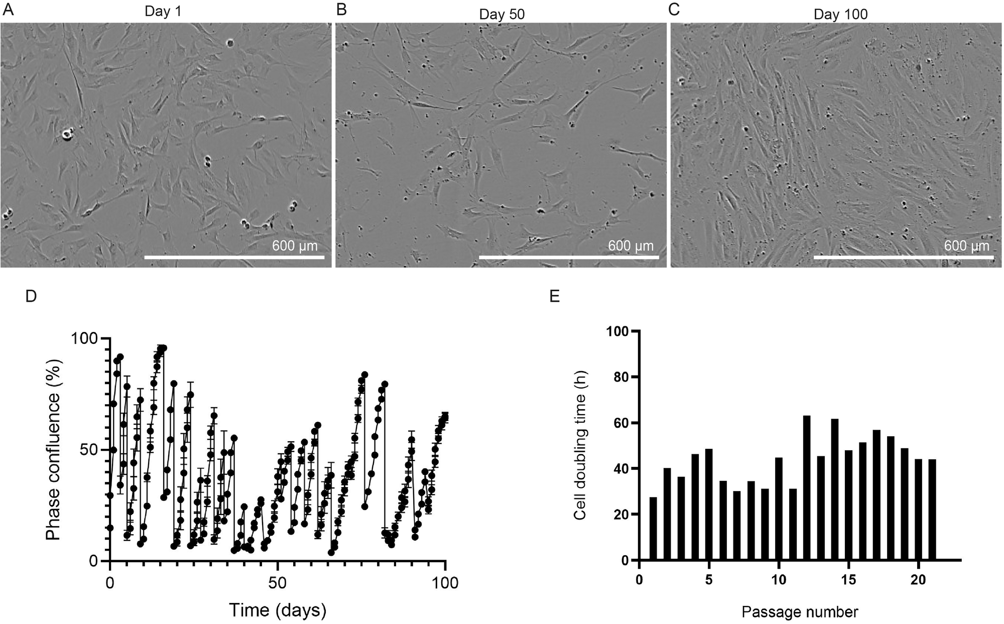
Phase contrast images of the isolated cells following A 1, B 50 or C 100 days of culture within an Incucyte S3 live cell imaging instrument. Scale bars = 600 μm. D Degree of cell confluence over 100 days of cell culture and 21 independent passages of the bovine preadipocytes. The degree of cell confluence, “phase confluence (%)”, was determined by the internal software of an Incucyte S3 live cell imager, which performs densitometry on phase contrast images acquired over the 100-day time course. E Cell doubling time over 21 independent passages, as calculated by the formula: doubling time = duration.ln(2) = duration x ln(2)/ln(final confluence/initial confluence). (Figure created using GraphPad Prism).
